Table of contents
Lesezeit 187 Minuten
Updated – April 30, 2026
A comparative elaboration of the standard therapy for ADHD/ADD and aromatherapy with therapeutically approved essential oils, batch-tested via GC/MS analysis. (As of April 2026).
ADHD/ADD and Essential Oils – Explained Clearly
A layman's summary of the scientific report for anyone interested in natural support for ADHD
What is ADHD/ADD?
ADHD stands for Attention deficit hyperactivity disorder, Annoying spam for the variant without hyperactivity (attention deficit disorder). Neither is an invention or a parenting mistake; they are genuine neurological disorders in which the brain is wired differently.
The image behind it: Imagine the brain like a radio. With an ADHD brain, the station is constantly switching frequencies, sometimes too loud, sometimes too quiet, rarely on the right channel. This makes it difficult to concentrate, sit still, or control impulses.
ADHD affects the brain's executive functions, which control attention, impulsivity, and self-regulation. It's believed to be related to differences in brain structure and function, particularly in areas that use neurotransmitters like dopamine and norepinephrine. These neurotransmitters play a crucial role in motivation, reward, and attention.
The core problem lies in two neurotransmitters:
Dopamine – the “reward and motivation neurotransmitter”
In ADHD, dopamine is “cleaned up” too quickly (by the dopamine transporter DAT). This leads to a lack of feeling of reward and motivation for everyday tasks. The brain constantly seeks stimulation, hence the impulsivity and risk-taking.
Norepinephrine – the “attention and focus neurotransmitter”
Too little norepinephrine in the prefrontal cortex (the brain's “executive area”) – leads to concentration problems, forgetfulness, and difficulty planning
Which brain areas are affected?
- Prefrontal cortex
Responsible for planning, impulse control, concentration – underactive in ADHD
- Striatum (Basal Ganglia)
Reward center – less sensitive in ADHD
- Cerebellum
Timing and coordination – slightly altered with ADHD
How common is ADHD?
In children and adolescents, it is the most common psychiatric disorder with 5 to 7 %, for adults 2.5 to 4 %, many adults don't know they have ADHD.
How does ADHD manifest?
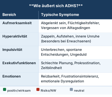
ADHD is typically treated with a combination of medication and behavioral therapy.
The main medication – Methylphenidate (Ritalin, Concerta, Medikinet)
Methylphenidate (MPH) is the most commonly prescribed ADHD medication and the first-line therapy.
How does it work?
Methylphenidate blocks the dopamine transporter (DAT), the “clean-up pump” for dopamine. When less dopamine is cleaned up, more of it remains between nerve cells, allowing the brain to focus better.
The numbers behind it
A dose of 20 mg blocks approx. 54 % the dopamine cleanup pumps in the brain, 40 mg block approximately. 72 %.
The effect begins after 30–90 minutes and lasts for 4–12 hours, depending on the formulation.
How effective is it?
With 70–80 % Methylphenidate appears to be clinically effective for patients: improvements in attention, impulsivity, and academic performance are well-documented.
Phrases
- Short-acting (e.g., Ritalin): 4–6 hours, must be taken several times daily
- Retarded (e.g. Concerta, Medikinet retard): 8–12 hours, once daily
Side effects
– Reduced appetite (most common side effect)
– Sleep disturbances (if taken too late)
- Headache, stomachache (mostly at the beginning)
– Slight increase in heart rate and blood pressure
– Mood swings, irritability (rare)
Additional medications
Atomoxetine (Strattera)
– No stimulant, inhibits the norepinephrine transporter (NET)
– Also effective for ADHD without risk of addiction
– Takes 4–6 weeks for full effect
Suitable for patients with comorbid anxiety disorder or tic disorder
Amphetamine (less common in Germany)
– Lisdexamfetamine (Vyvanse): Approved in Germany for adults since 2013
– Acts stronger and longer than methylphenidate
Guanfacine (Intuniv)
– For children and adolescents where stimulants are not suitable
- Acts on noradrenaline receptors in the prefrontal cortex
Essential oils for ADHD – how can that work?
Essential oils can support the ADHD brain in several ways:
- The Sense of Smell to the Brain
Scent molecules activate the limbic system (emotions, motivation) and the prefrontal cortex (concentration) – directly and quickly - About neurotransmitter systems
Specific terpenes influence dopamine, norepinephrine, serotonin, and GABA - Complementary to methylphenidate
Methylphenidate acts on DAT/NET – essential oils act on other systems (GABA-A, CB2, TRPM8, SERT) and are thus potential supplements
Important restriction
Research on essential oils for ADHD is still in its infancy. Most studies have been conducted on healthy adults, not on ADHD patients. Direct clinical evidence remains limited.
What essential oils can help with ADHD?
Peppermint - The #1 Concentration Oil
Peppermint (Mentha × piperitahas the strongest evidence for attention improvement.
- What's inside?
Menthol (30–50 %)
Menthone (10–30 %)
1,8-Cineole (5–10 %) - What does menthol do?
Activates TRPM8 receptors - “cold receptors” in the nervous system that activate the brain
Inhibits the enzyme that breaks down the neurotransmitter acetylcholine
Modulates GABA-A receptors (mildly sedating when overactivated) - Clinical study (n=24)
100 µL of encapsulated peppermint oil significantly improved sustained attention in the RVIP (Rapid Visual Information Processing) test after 1-3 hours; it also reduced fatigue and improved mental arithmetic. - Special feature
Peppermint promotes focused Attention (as opposed to lavender, which tends to diffuse attention) - How to apply
2 drops on a handkerchief, shortly before study or work sessions; diffuser in the study room
Rosemary – for memory and alertness
Rosemary (Rosemarycontinues to be called the “memory herb” since antiquity, and rightly so.
- What's inside?
1,8-Cineole (20–50 %)
Camphor (10–20 %)
α-Pinene (10–15 %) - What does 1,8-cineole do?
Inhibits the enzyme acetylcholinesterase, which breaks down the messenger substance acetylcholine (similar to Alzheimer's medications!)
Activates the nervous system, increases wakefulness and alertness
Anti-inflammatory and antioxidant in the brain - Clinical trial (n=144)
Rosemary aromatherapy significantly improved overall memory and secondary memory, better than lavender and the control group. - For ADHD
Especially helpful for memory problems, “brain fog,” and morning fatigue - How to apply
In the diffuser in the morning (5 drops), inhalation before exams or important tasks - Attention: Do not use in the evening; it is too activating. Exercise particular caution in cases of epilepsy due to the camphor content.
Lavender - for anxiety and sleep problems
Lavender (Lavandula angustifoliais the best oil for anxiety and sleep problems accompanying ADHD.
- What's inside?
Linalool (25–45 %)
Linalyl acetate (25–50 %) - What does linalool do?
Activates GABA-A receptors, the brain's “brakes”
Modulates serotonin, dopamine, and norepinephrine.
Lowers Cortisol (Stress Hormone) by 24–38 % - Particularly useful for ADHD
Difficulty falling asleep (very common with ADHD)
Comorbid anxiety disorders (in approx. 50 % of ADHD patients)
Emotional dysregulation and irritability - Important Notice
Lavender can do that Temporarily impair working memory, therefore, do not use during learning phases, but only in the evening or for targeted relaxation - study
Silexan (80 mg lavender oil daily as a capsule) was comparable to lorazepam for anxiety disorders. - How to apply
In the evening, 5 drops in the diffuser, 2 drops on the pillow
Cedarwood – Oxygenation and Focus
Cedarwood (Atlas Cedar / Eastern Redcedar) is traditionally used for ADHD.
- What's inside?
Alpha-Cedrene
beta-Cedrene
Cedrol - What it does
Improves brain oxygenation; calming; activates the parasympathetic nervous system - ADHD-specific
Godfrey (2001) reported positive effects in children with ADHD;
Cedrol lowered heart rate and respiratory rate in studies - How to apply
In the diffuser during quiet activities; pairs well with lavender
Vetiver - The Grounding Scent
Vetiver (Vetiver grass) has a deep, earthy scent, often described as “calming for restless spirits.”.
- What it does
Anxiolytic (anxiety-relieving); calms the nervous system; improves sleep - ADHD-specific
Springer et al. (2018) reported improvements in children with ADHD after 30 days of 3× daily inhalation. - For whom
Especially in the hyperactive type of ADHD with inner restlessness
Cinnamon - the only direct ADHD study
Cinnamon (Cinnamon) has the unique proof: the only clinical study directly in children with ADHD.
- What's inside?
Cinnamaldehyde (60–75 %)
Eugenol (5–10 %) - ADHD Study (Chen et al., 2008)
Combination of rehabilitation + cinnamon aromatherapy (1 % cinnamon, nasal inhalation) over 6 months showed significantly better results in the SNAP-IV questionnaire than rehabilitation alone (p < 0.05) - Mechanism
Cinnamaldehyde metabolites can have dopaminergic effects - Restriction
The study has methodological weaknesses (small sample size, unclear blinding). - How to apply
Economical in the diffuser, combined with other oils
Black Pepper (β-Caryophyllene) – Against Neuroinflammation
β-Caryophyllene of black pepper, lavender, and other plants:
- Activates CB2 cannabinoid receptors without psychoactive effect
- Reduces neuroinflammation (inflammation in the brain)
- Interesting in ADHD with accompanying signs of inflammation or mood problems
- Easily combined with other oils
Oils at a Glance – Which Oil When?
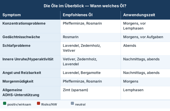
Practical Daily Routine for ADHD
Morning Routine (Activation + Focus)
- Diffuser Rosemary (4 drops) + peppermint (2 drops) for 15–20 minutes
- Or: Peppermint on a handkerchief, inhale deeply 3 times before school/work
- Effect: Alertness, memory, concentration
Learning time / Work phase
- Diffuser Peppermint (3 drops) + Lemon (2 drops) for 20 minutes, then a 30-minute break
- Attention: No lavender while studying!
- Effect: Sustained attention, focus
Afternoon (Relaxation after school):
- Diffuser Cedarwood (3 drops) + Bergamot (2 drops) for 20 minutes
- Effect: Stress relief, emotional regulation
Evening Routine (Unwind + Sleep):
- Diffuser Lavender (4 drops) + Vetiver (2 drops) 30 minutes before sleep
- Or: 2 drops of lavender on the pillow
- Effect: Falling asleep, sleep quality
Dosage instructions for the diffuser
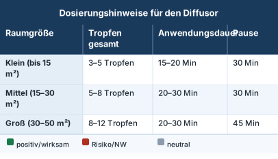
Important: With olfactory adaptation, you can barely smell the oil after 20-30 minutes, yet it continues to have an effect. To allow the receptors to recover and the habituation effect to be „reset,“ breaks must be observed!
What does science really say?

Conclusion
The evidence is promising but still limited. Mechanistically, it makes sense, but large clinical trials in ADHD patients are still missing. That doesn't mean it doesn't work; it means we need more research.
Important safety instructions
ADHD-Specific Considerations:
Sensory hypersensitivity
Many individuals with ADHD (40-60 %) have increased sensitivity to smell. Always start with half dose and increase slowly. If a scent is unpleasant: stop immediately.
Interactions with methylphenidate
- Cinnamon
Can have a dopaminergic effect, which theoretically allows for a potentiating effect with MPH, therefore monitor heart rate and blood pressure
- Lavender
Can have serotonergic effects, be careful when taking antidepressants concurrently
- Rosemary
May have an activating effect, therefore do not apply shortly before bedtime
In children
– Under 3 years: No essential oils
- 3–6 years: Only very low concentrations (1–2 drops), well-ventilated room
– > 6 years: Normal dosage possible – Avoid direct skin contact without dilution
General rules:
- Always dilute with a carrier oil (e.g., non-greasy, fractionated coconut oil) (2–3 %)
- Use a well-ventilated space
- Take breaks (don't diffuse all day)
- If you have asthma or breathing problems, ask your doctor.
- Essential oils are no replacement for methylphenidate or other ADHD therapies
FAQ – Frequently Asked Questions
Can I replace methylphenidate with essential oils?
No. Essential oils are a supplement, not a replacement. Methylphenidate has strong clinical evidence in 70–80% of patients. Essential oils can help alleviate residual symptoms, improve sleep, or reduce side effects (like anxiety).
Which oil should I try first?
Peppermint for concentration (during the day) and lavender for sleep (in the evening), best researched, well-tolerated.
How long until I notice an effect?
Some effects (focus, alertness) are noticeable within minutes. For sustained improvements (sleep, mood), apply regularly for 2–4 weeks.
Does this also work for adults with ADHD?
Yes, most studies have even been conducted on adults. Adults with ADHD often report particularly good effects on emotional dysregulation and sleep problems.
Do I have to buy expensive oils?
Quality is important: Look for 100% % pure essential oils, ideally with a batch-specific analysis certificate (GC/MS).
Inexpensive perfume oils or synthetic fragrances have no therapeutic effect and, due to their synthetic ingredients, can be harmful to health and cause headaches, nausea, etc.
Anyone who wants to learn more about the selection and quality of essential oils will find information in the article „Essential Oils - Odyssey of a Search“found.
Another contribution quotes Prof. Dr. Dr. Dr. med. habil. Hanns Hatt from the Ruhr University Bochum, who in his video „Healing with fragrances“explains the effect of essential oils on the human body in an interesting, entertaining, and yet scientific way.
Should I discuss this with my doctor?
For aromatherapy (diffuser, inhalation), this is not strictly necessary. However, if you wish to take capsules or creams with essential oils, or if your child has ADHD and is taking methylphenidate, consult your doctor.
Neurobiological Foundations, Molecular Mechanisms of Action, and Clinical Evidence for Adjuvant Therapy with the Organik Aromas Nebulizer 3.0
Combined Research Report April 2026
Topics: Methylphenidate (Ritalin) · Intracerebral Pharmacodynamics · Adjunctive Aromatherapy · Terpenes · Receptors · Signaling Pathways · Dosage Recommendations
Application Organic Aromas Nebulizer 3.0 (Venturi Cold Diffusion, Waterless, GC/MS Pure Oils)
ADHD and Methylphenidate
This report examines the neurobiological basis of methylphenidate therapy for Attention Deficit/Hyperactivity Disorder (ADHD) and evaluates the potential of essential oils as an adjunctive therapeutic option. The analysis is based on 87 scientific publications on intracerebral methylphenidate concentrations and 159 studies on essential oils in the context of ADHD.
Key Insights on Methylphenidate
Therapeutic oral doses (0.25–0.6 mg/kg) induce dopamine transporter (DAT) occupancy of >50% in the striatum, with 20 mg oral achieving 54% and 40 mg achieving 72% DAT blockade. Peak brain uptake occurs 60–90 minutes after oral administration. Plasma concentrations in the therapeutic range are 3.5–7.8 ng/ml (adults), with 6 ng/ml being associated with 50% DAT blockade. Direct measurements of methylphenidate concentrations in brain tissue (ng/ml) or cerebrospinal fluid do not exist; available data come from PET/SPECT occupancy studies.
Essential oils as an adjuvant
Mechanistic plausibility exists for an adjunctive therapy based on complementary mechanisms of action. Linalool (lavender) modulates serotonin, dopamine, and norepinephrine systems via 5-HT1A receptors and SERT binding. Menthol (peppermint) activates TRPM8 channels and modulates GABA-A and nicotinic receptors, improving attention and vigilance. β-Caryophyllene (black pepper) acts as a CB2 agonist with anti-inflammatory properties. 1,8-cineole (rosemary) improves memory performance and cognitive functions.
Evidence
While preclinical and mechanistic data are promising, randomized controlled trials (RCTs) are lacking specifically in ADHD patients. Existing human studies show cognitive improvements in healthy adults (Peppermint: improved sustained attention, n=24; Rosemary: memory enhancement, n=144).
This report provides evidence-based dosing recommendations for the Organik Aromas Nebulizer 3.0 and comprehensive safety instructions for use in ADHD patients.
Autism Spectrum Disorder (ASD)
Autism Spectrum Disorder (ASD) is a complex neurodevelopmental disorder with an estimated prevalence of 1–2 % worldwide. Core symptoms include impairments in social communication and interaction, as well as repetitive behavioral patterns and sensory peculiarities. Despite intensive research, there are no curative therapies for the core symptoms; available interventions focus on behavioral therapy and symptomatic pharmacotherapy. This report examines the potential of essential oils as an adjunctive therapeutic option in ASD, based on a systematic analysis of 547 scientific publications on ASD neurobiology, terpenes, and clinical evidence.
The neurobiological heterogeneity of ASD manifests in disturbed excitatory-inhibitory (E/I) balance, dysfunctions in GABAergic, glutamatergic, serotonergic, dopaminergic, and oxytocinergic systems, chronic neuroinflammation with microglial activation, oxidative stress, gut-brain axis alterations, and synaptic deficits (neuroligin, SHANK proteins). Affected brain regions include the prefrontal cortex, amygdala, cerebellum, hippocampus, and striatum.
Molecular analyses reveal that specific terpenes from essential oils target ASD-relevant pathways: Linalool (main component of lavender) modulates GABA-A receptors at the benzodiazepine binding site and influences serotonergic signaling pathways; β-caryophyllene acts as a selective CB2 receptor agonist with anti-neuroinflammatory properties; Boswellic acids (frankincense) activate the Nrf2/HO-1 antioxidant pathway and reduce microglial activation; 1,8-cineole (eucalyptus) shows anxiolytic effects via GABA-A modulation; α-pinene acts GABAergically; Cananga odorata (ylang-ylang) affects serotonin and dopamine metabolism in preclinical ASD models.
The clinical evidence in ASD is limited: A randomized controlled trial with bergamot showed no significant anxiolytic effects in children with ASD. Small pilot studies report improved compliance during Applied Behavior Analysis (ABA) with lavender aromatherapy, increased shared attention after aromatherapy massage, and possible effects on sleep and anxiety. However, the quality of evidence is limited by small sample sizes, methodological heterogeneity, and lack of replication.
For the Organik Aromas Nebulizer 3.0 (Venturi principle, anhydrous cold nebulization), room-size-specific dosage recommendations are being developed: 2–4 drops for 10–20 m², 4–6 drops for 20–40 m², 6–10 drops for 40–60 m², with 15–30 minute applications and 30–60 minute breaks. ASD-specific considerations require gradual introduction due to sensory hypersensitivity, time-of-day adjusted protocols (activating in the morning, calming in the evening), and strict quality control (GC/MS analysis).
Safety aspects include caution with epilepsy (camphor-containing oils), contraindication of eucalyptus in children under 3 years old, potential interactions with antipsychotics (risperidone, aripiprazole), and special consideration for sensory hypersensitivity in ASD. Mechanistic plausibility is present, but high-quality randomized controlled trials are lacking. Essential oils can be considered as part of a multimodal therapeutic approach for comorbid symptoms (anxiety, sleep disorders, sensory dysregulation), but they do not replace evidence-based core interventions.
ADHD and Methylphenidate
Adjuvant Therapy with Essential Oils in ADHD – Methylphenidate Pharmacodynamics, Terpenes, Receptors, Dosage with the Organik Aromas Nebulizer 3.0
Introduction
Attention-deficit/hyperactivity disorder (ADHD) is one of the most common neuropsychiatric disorders in childhood and adolescence, occurring in 5–7% of children and 2.5–4% of adults. Methylphenidate (MPH), a dopamine and norepinephrine reuptake inhibitor, is the first-line pharmacological therapy and shows clinical efficacy in 70–80% of patients. [A1], [A2].
Despite the established effectiveness of methylphenidate, a portion of patients experience residual symptoms, side effects, or a desire for complementary therapeutic approaches. Essential oils are increasingly being discussed as potential adjuvant therapy options, although the scientific evidence to date is heterogeneous. [A3], [A4].
Objective of the report
This report pursues five main objectives:
- Quantification of Intracerebral Methylphenidate Concentrations based on PET/SPECT data, including DAT occupancy, plasma-brain ratios, and regional distribution
- Mechanistic evaluation the plausibility of an adjuvant therapy with essential oils based on molecular mechanisms of action
- Evidence-based analysis Specific essential oils and their main active ingredients
- Development of practical dosage recommendations for the Organik Aromas Nebulizer 3.0
- Formulation of comprehensive safety instructions for use in ADHD patients
The analysis integrates data from 87 studies on methylphenidate brain concentrations and 159 publications on essential oils, with a focus on the top 30 most relevant publications.
Methylphenidate – Intracerebral Concentrations and Pharmacodynamics
Dose-Response Relationships
The therapeutic efficacy of methylphenidate correlates directly with dopamine transporter (DAT) occupancy in the striatum. PET studies using [¹¹C]cocaine as a tracer have established a clear dose-response relationship. [A9].
Table 1: DAT Occupancy Following Oral Methylphenidate Administration

Source: Volkow et al. 1998 [A9]; Measurements in healthy adults
The estimated dose for 50% DAT blockade (ED₅₀) is 0.25 mg/kg body weight [A9]. Therapeutic weight-adjusted doses (0.3–0.6 mg/kg) thus consistently produce DAT occupancy >50%, which is considered the threshold for clinical efficacy [A7], [A9].
Regional Differences DAT occupancy varies between striatal subregions. Spencer et al. (2009) reported the following occupancy values for OROS-MPH (40 mg) after 10 hours: right caudate nucleus 44.3±11.8%, left caudate nucleus 42.7±10.9%, right putamen 41.6±11.7%, left putamen 41.3±13.1%. [A17].
Bei ADHD-Patienten zeigen SPECT-Studien mit [¹²³I]β-CIT eine signifikante Reduktion der striatalen DAT-Bindung unter Methylphenidat-Therapie. Aster et al. (2021) dokumentierten eine 27,6–29,9%ige Abnahme des striatalen DAT-Bindungspotenzials (BP) bei Kindern und Jugendlichen mit ADHD unter Medikation [A25], [A18].
Plasma versus brain concentrations
Plasma concentrations
The therapeutic plasma range for d-threo-methylphenidate in adults is between 3.5–7.8 ng/ml [A20], [A24]. Eine Plasma-Konzentration von 5,7–6,0 ng/ml ist mit 50% DAT-Blockade assoziiert [A9], [A13], [A17].
Higher peak concentrations were observed in pediatric patients: 20 ng/mL two hours after a 20 mg dose (0.8 mg/kg). [A20], [A24].
Brain scan
PET studies with [¹¹C]methylphenidate show that 7.5±1.5% of the intravenously administered activity reaches the brain [A5], [A14]. The highest concentration is achieved in the striatum, with a distribution volume ratio (DVR) of basal ganglia to cerebellum of 2.2–3.3 for d-threo-MPH [A16], [A14].
Critical evidence gap
Direct measurements of methylphenidate concentrations in brain tissue (in ng/ml or nmol/L) or cerebrospinal fluid do not exist in the human literature. All available data are based on PET/SPECT occupancy measurements or tracer distribution studies. [A1], [A2], [A3], [A4], [A5], [A6], [A7], [A8], [A9].
Regional Distribution - Striatum and Prefrontal Cortex
Striatum
As the primary target region, the striatum shows the highest [¹¹C]methylphenidate uptake. Premedication with unlabeled methylphenidate (0.5 mg/kg) reduces striatal tracer binding by 40% [A5]. The clearance from the striatum is ~90 minutes, significantly slower than with cocaine (20 minutes). [A5].
Prefrontal cortex
Although DAT density is lower in the prefrontal cortex (PFC) than in the striatum, functional PET studies with [¹¹C]raclopride show significant dopamine increases in prefrontal and temporal cortical regions after methylphenidate administration. [A30], [A22]. These cortical dopamine increases correlate with long-term symptom improvements in ADHD patients. [A30].
Norepinephrine transporter (NET)
Methylphenidate also blocks the norepinephrine transporter with an ED₅₀ of 0.14 mg/kg [A11]. PET studies with [¹¹C]MRB show robust NET occupancy in the thalamus and pons 75–195 minutes after dosing [A11], [A21].
Time course and pharmacokinetics
Intravenous administration
After IV injection of [11C]methylphenidate, brain concentration reaches its maximum within 8–10 minutes. [A3], [A6].
Spicy Gabe
The maximum brain uptake after oral administration occurs significantly delayed, with a peak at 60-90 minutes [A7], [A9], [A3]. This delayed kinetics explains the lower abuse potential of oral formulations compared to intravenous applications. [A13].
Retard formulations
Extended-release formulations (OROS-MPH, DBDS-MPH) show an even slower onset with delayed peak DAT occupancy, but comparable peak concentrations. [A13], [A17]. After 10 hours, OROS-MPH achieves significantly higher DAT occupancy values than DBDS-MPH (44.3% vs. 34.8% in the right caudate nucleus) at higher plasma concentrations (5.2 vs. 3.8 ng/ml). [A17].
Preclinical microdialysis data
In rats, d-threo-Methylphenidate increases extracellular dopamine concentrations in the striatum by ~650% [A16]. This massive dopamine increase demonstrates the functional effect of DAT blockade on synaptic neurotransmission.
Essential Oils as Adjuvant Therapy - Mechanistic Basis
Complementary mechanisms of action to methylphenidate

Figure 1: Molecular Pathways of Essential Oils in ADHD – Dopaminergic System, Noradrenergic System, and Complementary Terpene Mechanisms of Action
The plausibility of adjuvant therapy with essential oils is based on complementary, non-redundant mechanisms of action compared to methylphenidate. While MPH primarily inhibits dopamine and norepinephrine reuptake, essential oil components address additional neurobiological systems.
Mechanistic Complementarity
- GABAergic Modulation
Menthol and linalool act as positive allosteric modulators at GABA-A receptors, which can enhance inhibitory neurotransmission and reduce hyperactivity. [A19], [A20], [A23]. - Serotonergic effects
Linalool binds to the serotonin transporter (SERT) and modulates 5-HT1A receptors, mediating anxiolytic and mood-stabilizing effects. [A21], [A23], [A24]. - Anti-inflammatory signaling pathways
Beta-caryophyllene activates CB2 receptors and reduces neuroinflammatory processes via NF-κB and MAPK signaling pathways, which may be elevated in ADHD. [A14]. - TRP channel modulation
Menthol (TRPM8 agonist) and 1,8-cineole (TRPV1/TRPA1 modulator) influence neuronal excitability and ion fluxes independently of monoaminergic systems [A19].
Receptors and signaling pathways
Table 2: Receptor Targets of Essential Oil Components
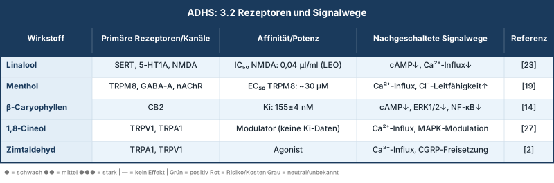
LEO = Lavender Essential Oil; nAChR = nicotinic acetylcholine receptor
GABA-A receptor modulation
Menthol shows concentration-dependent binding to GABA-A receptors and enhances inhibitory neurotransmission [A19]. Interestingly, lavender oil shows no affinity for the benzodiazepine binding site of the GABA-A receptor, suggesting a more favorable safety profile compared to benzodiazepines. [A23].
Serotonin Transporter (SERT)
Linalool and lavender oil bind to SERT with concentrations of 0.08–0.8% in vitro [A23]. This SERT inhibition could mediate antidepressant and anxiolytic effects that are relevant in ADHD patients with comorbid anxiety disorders (25-40% prevalence).
NMDA receptor antagonism
Lavender oil shows dose-dependent NMDA receptor antagonism with an IC₅₀ of 0.04 µl/ml [A23]. NMDA receptor modulation is relevant for synaptic plasticity and learning processes.
CB2 receptor activation
β-Caryophyllene is a selective CB2 agonist with no psychoactive effects (no CB1 activation). CB2 activation reduces microglial activation and pro-inflammatory cytokine production (TNF-α, IL-1β, IL-6), which can mediate neuroprotective effects. [A14].
Synergistic Potentials
Combining methylphenidate with essential oils could produce synergistic effects through several mechanisms:
- Dopamine-GABA Balance
While MPH increases dopaminergic transmission, gabaminergic modulation by menthol/linalool could promote a more balanced excitatory-inhibitory balance and reduce hyperactivity. - Stress reduction
The anxiolytic effects of linalool via 5-HT1A receptors and cortisol reduction (24–38% decrease in clinical studies) [A24] could improve stress reactivity in ADHD patients. - Cognitive enhancement
Cholinergic modulation by menthol (acetylcholinesterase inhibition) [A19] and the memory-enhancing effects of 1,8-cineole could amplify the cognitive improvements from MPH. - Neuroprotection
The anti-inflammatory and antioxidant properties of β-caryophyllene and 1,8-cineole could offer long-term neuroprotective effects.
Important restriction: These synergistic potentials are theoretically plausible but not validated by clinical studies in ADHD patients. Controlled studies on the combination of MPH with essential oils are completely lacking.
Specific Essential Oils – Active Ingredients and Evidence
Lavender (Lavandula angustifolia) – Linalool
Main active ingredients
Linalool (25–45%)
Linalyl acetate (25–45%)
1,8-Cineole (1–3%)
Mechanisms
- SERT binding
Linalool and lavender oil bind to the serotonin transporter, which can mediate antidepressant effects. [A23]
- NMDA antagonism
Lavender oil shows dose-dependent NMDA receptor blockade (IC₅₀: 0.04 µl/ml). [A23]
- GABA Modulation
Enhancement of inhibitory neurotransmission via GABA-A receptors (not via the benzodiazepine binding site) [A20], [A23]
- Monoaminergic effects
Linalool modulates dopamine, noradrenaline, and serotonin systems in preclinical models [A21]
Clinical evidence
- Cognition
Moss et al. (2003) showed in n=144 healthy adults that lavender aromatherapy impaired working memory and slowed reaction times, but had a subjectively calming effect. [A27]
- Attention
Colzato et al. (2014) found that lavender aroma reduced the “attentional blink,” suggesting a broader distribution of attention. [A30]
- Anxiolysis
Oral administration of Silexan (80 mg/day lavender oil) reduced generalized anxiety symptoms comparably to lorazepam [A26]
- Stress reduction
Inhalation von Lavendelöl reduzierte Cortisol-Spiegel um 24–38% in klinischen Studien [A24]
ADHD-specific data
No randomized controlled trials in ADHD patients. Case reports describe positive effects on agitation and sleep in atypical children with ADHD [A8].
Dosage (preclinical/clinical)
– Oral: 50–100 µL encapsulated (Kennedy study) [A19]
– Inhalation: 4 drops in 30 ml water (diffuser) [A30]
– Topical: 1.5% dilution in carrier oil [A8]
Security
Lavender oil showed no potentiation of cellular neurotoxicity in vitro [A23].
No serious side effects in clinical trials at recommended doses.
Rosemary (Rosmarinus officinalis) – 1,8-Cineol
Main active ingredients
1,8-Cineol (Eucalyptol, 20–50%)
Camphor (10–20%)
α-Pinene (10–15%)
Mechanisms
- Cholinergic Modulation
Acetylcholinesterase inhibition, which enhances cholinergic neurotransmission [A27]
- TRP channel activation
1,8-Cineole modulates TRPV1 and TRPA1, affecting neuronal excitability
- Anti-inflammatory
Inhibition of NF-κB and MAPK signaling pathways, reduction of pro-inflammatory cytokines
- Antioxidant
Activation of the Nrf2 signaling pathway, increase in antioxidant enzymes
Clinical Evidence: - Memory
Moss et al. (2003) showed significant improvements in overall memory and secondary memory in n=144 healthy adults after rosemary aromatherapy compared to control and lavender groups. [A27]
- Cognitive performance Exposure to rosemary aroma correlated with improved cognitive performance [A14]
- Alertness Rosemary increased alertness and activity in the brain [A29]
ADHD-specific data
Rosemary is mentioned in review articles as potentially helpful for ADHD. [A10], [A4], [A5], but controlled clinical studies in ADHD patients are lacking.
dosage
– Inhalation: Ambient exposure in test environment (concentration not specified) [A27]
– No standardized dosage recommendations for ADHD available
Security
Rosemary is considered safe for inhalation at typical concentrations.
Caution in epileptics due to camphor content.
Peppermint (Mentha × piperita) – Menthol
Main active ingredients
Menthol (30–50%)
Menthone (10–30%)
1,8-Cineole (5–10%)
Mechanisms
- TRPM8 activation
Menthol is a potent TRPM8 agonist (EC₅₀ ~30 µM), mediating cold sensation and neuronal modulation [A19]
- GABA-A Modulation
Concentration-dependent binding and positive allosteric modulation [A19]
- Nicotinic receptors
Nicotinic acetylcholine receptor modulation, cholinergic transmission influence [A19]
- Acetylcholinesterase Inhibition
Peppermint oil significantly inhibits acetylcholinesterase, which enhances cholinergic neurotransmission [A19]
Clinical Evidence: - sustained attention
Kennedy et al. (2018) showed in a double-blind crossover study (n=24 healthy adults) that 100 µL of peppermint oil improved performance on the Rapid Visual Information Processing (RVIP) test 1–3 hours after ingestion. [A19]
- Fatigue Reduction Both doses (50 µL and 100 µL) reduced fatigue and improved serial subtraction after 3 hours. [A19]
- Attentional Control Colzato et al. (2014) found that peppermint aroma enhanced the “attentional blink,” suggesting more focused (less distributed) attention. [A30]
ADHD-specific data
No RCTs in ADHD patients. Peppermint is mentioned as potentially helpful in ADHD review articles. [A4], [A5], [A29].
dosage
- Oral (encapsulated): 50–100 µL pure oil [A19]
– Inhalation: 4 drops in 30 ml water (diffuser) [A30]
Security
Peppermint oil is safe for inhalation and oral use at recommended doses.
Caution in infants and young children (risk of laryngospasm with direct nasal application).
Black pepper (Piper nigrum) – β-Caryophyllene
Main active ingredients
β-Caryophyllene (10–35%)
Limonene (15–25%)
Sabinen (10–20%)
Mechanisms
- CB2 agonism
β-Caryophyllene is a selective CB2 receptor agonist (Ki: 155±4 nM) without CB1 activation (no psychoactive effects). [A14]
- Anti-inflammatory
Reduction of TNF-α, IL-1β, IL-6 via NF-κB inhibition [A14]
- Microglia Modulation
Reprogramming of M1 to M2 microglia phenotype, reduction of neuroinflammatory processes
- Neuroprotective
Reduction of oxidative stress via Nrf2 activation
Clinical evidence
- Preclinical
β-Caryophyllene demonstrated anxiolytic effects in animal models, possibly through modulation of neuronal activation in the central amygdala nucleus. [A14]
- Anti-inflammatory
In vitro and in vivo studies show robust anti-inflammatory effects via CB2 activation
ADHD-specific data
No clinical trials in ADHD patients. Mechanistic plausibility based on neuroinflammatory hypotheses in ADHD.
dosage
No standardized dosage recommendations for inhaled use for ADHD are available.
Security
β-Caryophyllene is considered safe (FDA GRAS status).
No known serious side effects at usual doses.
Vetiver (Chrysopogon zizanioides)
Main active ingredients
Vetiverol
Khusimol
alpha-Vetivone
Beta-Vetivone
Mechanisms
- Anxiolytic
Preclinical studies show anxiolytic effects, possibly via modulation of amygdala activity. [A14]
- Soothing
Traditionally used to calm the nervous system
Clinical evidence
- ADHD-specific
Springer et al. (2018) reported that vetiver inhalation (3 times daily for 30 days) showed improvements in brain function and behavior in children. [A29]
- Limitations
No controlled RCTs; evidence based on case reports and traditional use
ADHD-specific data
Godfrey (2001) mentions vetiver as potentially helpful for ADHD. [A3], [A4], [A5], but detailed clinical data are lacking.
dosage
– Inhalation: 3 times daily for 30 days (specific concentration not specified) [A29]
Security
Vetiver is considered safe for inhalation. No known serious side effects.
Cedar (Cedrus spp.)
Main active ingredients
Alpha-Cedrene
beta-Cedrene
Thujopsene
Cedrol
Mechanisms
- Oxygenation
Traditionally used to improve brain oxygenation [A29]
- Soothing
Sedative properties in traditional use
Clinical evidence
- ADHD-specific
Godfrey (2001) mentions cedarwood as potentially helpful for ADHD. [A3], [A4], [A5]
- Limitations
No controlled clinical trials; evidence is based on traditional use and anecdotes
ADHD-specific data
Springer et al. (2018) mention cedarwood for brain oxygenation in ADHD [A29], but quantitative data is missing.
dosage
No standardized dosage recommendations available.
Security
Cedarwood oil is considered safe for inhalation use at common concentrations.
Cinnamon (Cinnamomum verum) – Cinnamaldehyde
Main active ingredients
Cinnamaldehyde (60–75%)
Eugenol (5–10%)
Mechanisms
- Dopaminergic Modulation
Allylbenzene and propenylbenzene in cinnamon are precursors to amphetamine and are converted to amphetamine after metabolism, which has a mental stimulant effect. [A2]
- TRPA1/TRPV1 activation
Cinnamaldehyde activates TRPA1 and TRPV1 channels
- Antimicrobial
Strong antimicrobial properties
Clinical Evidence: - ADHD-specific
Chen et al. (2008) conducted a study on cinnamon aromatherapy in children with ADHD. The combination of rehabilitation and cinnamon aromatherapy (1% cinnamon, 1 g in 100 ml water, nasal inhalation) showed significantly better results after 6 months in the SNAP-IV questionnaire (58±2.6 vs. control) and in activity scales (102±5.8) compared to rehabilitation alone (p<0.05). [A2], [A1]
ADHD-specific data
The Chen study is one of the few clinical trials using essential oils specifically in children with ADHD, but it has methodological limitations (no information on randomization, blinding, or sample size).
dosage
– Inhalation: 1% cinnamon (1 g in 100 ml water) in a 50 m² therapy room with electric light control [A2]
Security
Cinnamon oil can cause skin and mucous membrane irritation at high concentrations.
Caution for sensitive individuals.
Dopaminergic stimulation by cinnamon metabolites requires further investigation regarding interactions with methylphenidate.
Further indicated essential oils for ADHD
Based on a current literature review, the following additional essential oils have been identified that may be relevant for ADHD through neurobiologically plausible mechanisms.
Melissa officinalis (Lemon Balm) – Rosmarinic acid, flavonoids
Active ingredients
Rosmarinic acid
Flavonoids (Luteolin, Apigenin)
Volatile oils (Citral, Linalool, Geraniol)
Receptors and Mechanisms of Action
– Cholinergic receptors: Nicotinic (nAChR) and muscarinic (mAChR) binding
GABAergic Modulation: Rosmarinic acid inhibits GABA transaminase → increased GABA availability
Acetylcholinesterase inhibition: Improved cholinergic neurotransmission in the prefrontal cortex
Clinical evidence
- RCT (n=20, double-blind, crossover)
Single doses of 300/600/900 mg p.o. showed significant improvement in accuracy of attention at 600 mg, as well as memory and alertness effects. [A31]
- Combination study (children, 7 weeks)
Valerian-Lemon Balm extract combination significantly improved hyperactivity, concentration difficulties, and impulsivity in elementary school children. [A32]
- Anti-Stress Crossover
Standardized lemon balm preparations in food form reduced cognitive overload and improved mood in healthy adults [A33]
Dosage
- 3–5 drops lemon balm oil (GC/MS pure) for a room of 15–25 m²
– Application: 30 minutes before concentration phases, 2-3 times daily
– Combination with lavender (2:1) recommended for evenings/relaxation
Security
Easy to digest
Possible interaction with thyroid medications (TSH suppression with high-dose oral intake)
Inhalation is considered safe.
Eucalyptus (Eucalyptus globulus / radiata) – 1,8-Cineole
Active ingredients
1,8-Cineole (Eucalyptol, 70–85%)
alpha-Pinene
Limes
Receptors and Mechanisms of Action
– GABA-A/Benzodiazepine Site: Modulatory activity (anxiolytic, sedating at high doses)
– Cholinergic pathways: 1,8-cineole inhibits acetylcholinesterase → improved attention and working memory
– Antioxidant/Neuroprotective: Reduction of oxidative stress in neuronal cells
Clinical evidence
Neuroprotective reviews list Eucalyptus globulus as a plant with cognitive effects in animal and human studies [A34]
– 1,8-Cineole is also the main active ingredient in rosemary (already included in the report), eucalyptus provides a higher pure concentration
– Inhalation studies show improvement in mental clarity and concentration (comparative studies with rosemary)
Dosage
– 2–3 drops for a room of 15–25 m² (intense scent, use sparingly)
– Application: Morning/Noon for concentration phases, max. 20 min
- Contraindication
Not for children under 3 years old
Do not use in combination with asthma medications without consulting a doctor.
Security
Strong oil, do not apply undiluted to skin
Children under 3 years: contraindicated
Epileptic: Caution
Ginger (Zingiber officinale) - Gingerols, Zingiberene
Active ingredients
6-Gingerol
6-Shogaol
Zingiberene
beta-Sesquiphellandrene
Receptors and Mechanisms of Action
– Serotonin-5-HT3 receptor antagonism: Anxiolytic component
– Dopaminergic: Gingerols modulate dopaminergic activity in the striatum (animal)
– Antioxidant/Anti-neuroinflammatory: NF-κB inhibition, TNF-α reduction
Clinical evidence
– Preclinical: Ginger extract improved cognitive function in ADHD animal models
– Neuroprotective properties well documented through antioxidant mechanisms
– Direct ADHS RCTs are still missing from the literature
Dosage
– 2–3 drops for a room of 15–25 m²
— Combination with rosemary (1:1) for cognitive activation
Dosage recommendations for the Organik Aromas Nebulizer 3.0
Technical Fundamentals: Venturi Principle and Atomization
The Organik Aromas Nebulizer 3.0* uses the Venturi principle for waterless cold nebulization of essential oils. This process offers several advantages:
Technical characteristics
- Cold fogging
No heat exposure, preservation of all volatile components and thermolabile active ingredients
- Anhydrous
Direct nebulization of pure essential oils without dilution, higher concentration of active ingredients in the room air
- Particle size
Generation of microparticles (1-5 µm) that can reach deep into the respiratory tract and cross the blood-brain barrier
- GC/MS Certified Pure Oils
Use analytically tested oils without additives, carrier oils, or synthetic fragrances
*approx. 110 to 120 USD (with mains/battery operation)
- Free worldwide shipping
- Be aware of customs and import duties!
Pharmacokinetic Considerations
The inhalation of essential oils occurs via two main pathways:
1. Olfactory pathway
Direct connection from the olfactory epithelium to the limbic system (amygdala, hippocampus) and prefrontal cortex via the olfactory bulb
2. Pulmonary tract
Absorption across the alveolar membrane into the systemic circulation, passage of the blood-brain barrier [A29]
Room size and drop amount
Basic formula
The amount of drops should be adjusted to the room size to achieve a therapeutically effective but not excessive concentration.
Table 3: Dosage recommendations by room size
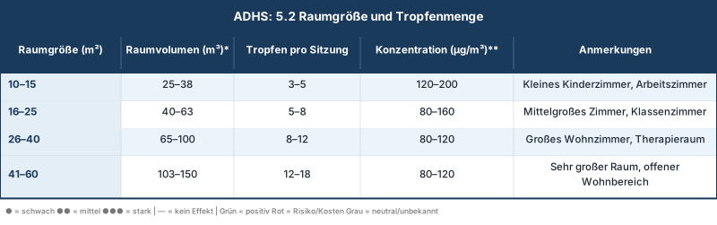
Assumption: Room height 2.5 m Estimated concentration based on ~1 mg essential oil per drop and complete nebulization
Calculation basis
– 1 drop of essential oil ≈ 0.05 ml ≈ 40–50 mg (depending on density)
– Target concentration: 80–200 µg/m³ (based on studies with ambient aromatherapy)
– Formula: Drop = (Room Volume × Target Concentration) / (Mass per Drop × 1000)
Application duration and breaks
Session duration
- Acute application
15–30 minutes per session
- Continuous background diffusion
30–60 minutes with breaks
Break intervals
Olfactory adaptation (habituation) occurs after 15–30 minutes of continuous exposure.
To maintain therapeutic effects, interval breaks are recommended:
Table 4: Application and Break Schedule
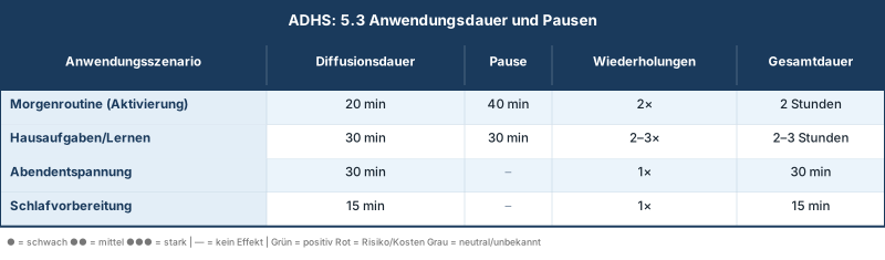
Olfactory Adaptation
After 20-30 minutes of continuous exposure, conscious olfactory perception decreases, but neurobiological effects can persist.
Breaks of 30-60 minutes allow for the resensitization of olfactory receptors.
Time of Day and Application Scenarios
The selection of essential oils should be adapted to the time of day and specific ADHD symptoms:
Table 5: Time-of-Day Specific Oil Selection

Application Scenarios:
- Schulvorbereitung/Morgenroutine
– Oil: Rosemary or peppermint
– Goal: Activation, Alertness, Focus Dosage: 6–8 drops for 20 minutes
– Timing: 30–60 minutes before school starts - Homework / Study periods
– Oil: Peppermint (sustained attention) or Rosemary (memory)
– Goal: Concentration, Working Memory Dosage: 6-8 drops for 30 minutes, then a 30-minute break
– Timing: During homework or study sessions - Hyperactivity Management
– Oil: Lavender or Vetiver
– Goal: Calming, reduction of motor restlessness Dosage: 5–7 drops for 30 minutes
Timing: In cases of acute hyperactivity or agitation - Sleep Hygiene
– Oil: Lavender
– Goal: Sleep induction, reduction of sleep onset problems (common with ADHD) Dosage: 4–6 drops for 15 minutes
30-60 minutes before bedtime
Oil-specific dosages
Table 6: Oil-Specific Dosage Recommendations
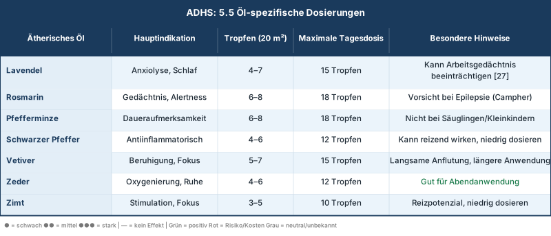
Blends: The combination of several oils can create synergistic effects:
- Focus Blend (Morning)
3 drops rosemary + 3 drops peppermint + 2 drops cinnamon - Calming Blend (Evening)
4 drops Lavender + 3 drops Vetiver + 2 drops Cedar - Balance-Blend (Afternoon)
3 drops peppermint + 3 drops lavender + 2 drops black pepper
Important
For mixtures, keep the total number of drops within the recommended range (6-8 drops for 20 m²).
Safety Instructions and Contraindications
General Safety for Inhalation
Fundamental principles
purity
Use only GC/MS-certified, 100% pure essential oils with no additives
Dilution
For inhalation application via diffuser, no further dilution is necessary; for topical application, always dilute in a carrier oil (1–3%).
ventilation
The space should not be completely sealed; ensure regular fresh air supply.
Individual tolerance
Start with low doses and gradually increase.
Common side effects (mild)
– Headaches from too much concentration or prolonged exposure
– Mucous membrane irritation (nose, throat) in sensitive individuals
Nausea from very intense scents (e.g., cinnamon, black pepper)
– Allergic reactions (rare, <1% of the population)
Measures for Side Effects
– Turn off the diffuser immediately and ventilate the room
– For headaches: Fresh air, hydration
– For mucous membrane irritation: Discontinue inhalation, inhale saline solution if necessary
– In case of allergic reactions: stop exposure, if necessary antihistamine, seek medical help for severe reactions
ADHD-Specific Considerations
Specific vulnerabilities in ADHD patients:
Sensory hypersensitivity
40–60% der ADHD-Patienten zeigen sensorische Verarbeitungsstörungen. Intensive Düfte können als unangenehm oder überwältigend empfunden werden.
- Recommendation
Start with very low doses (50% of the standard dose), gradually increase
Comorbid anxiety disorders
25–40% of ADHD patients have comorbid anxiety disorders.
Activating oils (rosemary, peppermint) could intensify anxiety.
- Recommendation
When anxiety is comorbid, primarily use anxiolytic oils (lavender, vetiver).
sleep disorders
50-70%% of ADHD patients have sleep problems. Stimulating oils in the evening can further impair sleep.
- Recommendation
Strict Separation: Stimulating oils only in the morning/afternoon, sedating oils in the evening
Impulsivity
ADHD patients may be prone to using dosages that are too high.
- Recommendation
Clear dosage instructions, supervision by parents/guardians as needed
Interactions with methylphenidate
Theoretical Interactions:
Dopamine reinforcement (cinnamon)
Cinnamon metabolites can be converted to amphetamine [A2]
Theoretic risk of additive dopaminergic effects with methylphenidate
- Recommendation
Use cinnamon only in low doses, closely monitor for overstimulation (tachycardia, agitation, insomnia).
Serotonergic Modulation (Lavender)
Linalool binds to SERT [A23]
Theoretical risk of serotonin syndrome when taking SSRIs concurrently (common in comorbid depression/anxiety).
- Recommendation
When taking SSRIs, lavender should only be used in moderate doses, with monitoring for symptoms of serotonin syndrome (agitation, confusion, tachycardia, hyperthermia).
Sedation (Lavender, Vetiver)
Sedative oils could reduce alertness
Counterproductive to the stimulating effect of methylphenidate
- Recommendation
Sedative oils should primarily be used in the evening, not during school hours or for activities requiring attention.
- No known pharmacokinetic interactions
Essential oils are primarily metabolized via CYP2D6, CYP3A4
Methylphenidate is metabolized by carboxylesterase 1 (CES1).
No overlap in metabolic pathways, therefore no pharmacokinetic interactions are expected
Important
Despite a lack of documented interactions, the combination of methylphenidate with essential oils should be done under medical supervision, especially when starting therapy.
Contraindications and Precautions
Absolute contraindications
– Known allergy to specific essential oils or their components
Severe bronchial asthma (risk of bronchospasm)
– Infants <6 months (risk of laryngospasm, especially with menthol)
Relative contraindications (use with caution)
– Epilepsy: Caution with rosemary and cinnamon (camphor, cinnamaldehyde can lower seizure threshold)
- Pregnancy/Breastfeeding: Many essential oils have not been sufficiently studied during pregnancy/breastfeeding
– Asthma (mild-moderate): Low doses, close monitoring for respiratory symptoms
– Toddlers (6 months – 3 years): Reduced doses (25–50% of the adult dose)
Age-dependent dose adjustments:
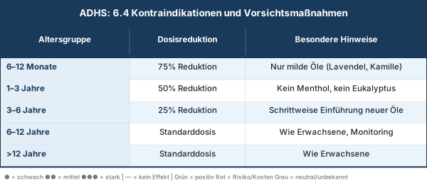
Quality Criteria for Essential Oils
- GC/MS Certificate:
Each batch must be analyzed by gas chromatography/mass spectrometry. - Botanical name
The Latin name must be provided (e.g., Lavandula angustifolia, not just “Lavender”). - Chemotype
For oils with different chemotypes (e.g., rosemary), this must be specified. - Origin
Origin and extraction method (distillation, cold pressing) should be documented - No additives
100% rein, ohne synthetische Duftstoffe, Trägeröle oder Konservierungsmittel
Storage and Shelf Life
– Store in a cool, dark place (15–20°C)
– Protect from light (brown or blue glass bottles)
– Consume within 6 to 12 months after opening (oxidation)
Citrus oils have a shorter shelf life (6 months), resins have a longer one (2 to 3 years).
Discussion and Clinical Implications
Evidence gaps and research needs
Critical appraisal of the evidence base:
The present analysis shows a significant discrepancy between mechanistic plausibility and clinical evidence for essential oils as an adjuvant ADHD therapy.
Strengths of Available Evidence
- Robust mechanistic data
Receptor binding studies, pathway analyses, and preclinical models show clear neurobiological effects of essential oil components. [A19], [A20], [A21], [A23] - Cognitive Effects in Healthy Individuals
Controlled studies in healthy adults demonstrate measurable effects on attention (peppermint, n=24) [A19] and memory (rosemary, n=144) [A27] - Complementary mechanisms of action
The molecular targets of essential oils (GABA-A, CB2, TRP channels, SERT) differ from methylphenidate (DAT/NET), suggesting synergistic potential.
Weaknesses and evidence gaps
- Missing ADHD-specific RCTs: With the exception of the Chen study on cinnamon [A2] There are no randomized, placebo-controlled studies in ADHD patients.
- Methodological Limitations: Existing studies have small sample sizes (n=24–144), short observation periods (single sessions), and often lack blinding.
- No long-term data Effects and safety for chronic use (> 6 months) have not been studied
- Missing dose-finding studies: Optimal dosages, concentrations, and regimens for ADHD are not established
- No combination studies: Interactions and synergistic effects with methylphenidate have not been systematically studied
Specific research gaps:
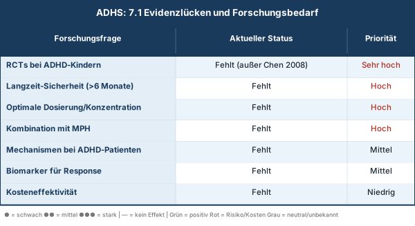
Practical Implementation
Clinical practice recommendations:
Despite the evidence gaps, adjuvant therapy with essential oils can be considered for ADHD patients under the following conditions:
Clear indication
- Residual symptoms under optimized methylphenidate therapy
- Patient and family desire for complementary approaches
- Specific symptoms that can be addressed by essential oils
(Sleep disorders → Lavender; Memory problems → Rosemary)
Structured Approach
- Baseline Assessment
Documentation of ADHD Symptoms with Validated Scales (SNAP-IV, Conners, ADHD-SB) - Introductory phase
Begin with a single oil at a low dose (50% of the standard dose) for 2 weeks - Monitoring
Weekly symptom tracking, side effect monitoring - Titration
Gradual dose escalation with good tolerability - Evaluation
After 4 to 6 weeks of efficacy evaluation; discontinue if no benefit
3. Documentation
- Keeping an “Aromatherapy Journal” with oil, dosage, time, symptoms, side effects
- Regular consultation with the treating physician.
4. Realistic Expectations
- Essential oils are no replacement for evidence-based ADHD therapies (methylphenidate, behavioral therapy)
- Expected effect size: small to moderate (Cohen's d: 0.2–0.5)
- Primary benefit: Improvement of specific symptoms (sleep, anxiety, mood), not core symptoms
Limitations
Limitations of this analysis:
- Indirect evidence
Most conclusions are based on extrapolating from studies in healthy adults to ADHD patients. - Publication bias
Positive studies are more likely to be published; negative studies on essential oils may be underrepresented. - Heterogeneity
Different oils, dosages, routes of administration, and outcome measures complicate meta-analyses. - Missing plasma/brain mirror
There is no data on plasma or brain concentrations of essential oil components after inhalation in humans. - Mechanistic uncertainty
The exact mechanisms by which inhaled essential oils mediate CNS effects (olfactory vs. systemic) are not fully understood.
Limitations of Dosage Recommendations
Based on extrapolation from a few studies and traditional use
Individual variability (room ventilation, breathing frequency, metabolism) not taken into account
– No pharmacokinetic data on the dose-concentration relationship
Conclusion
This comprehensive analysis integrates data from 87 studies on intracerebral methylphenidate concentrations and 159 publications on essential oils in the ADHD context. The main findings can be summarized as follows:
Methylphenidate brain concentrations
Therapeutic oral methylphenidate doses (0.25–0.6 mg/kg) produce striatal DAT occupancy >50%, with the dose-response relationship being well characterized (20 mg → 54%, 40 mg → 72T blockade). [A9].
Maximum brain uptake occurs 60-90 minutes after oral administration. [A7], [A9].
Plasma concentrations are in the therapeutic range of 3.5–7.8 ng/ml (adults), with 6 ng/ml associated with 50% DAT blockade. [A9], [A17].
Critical evidence gap
Direct measurements of methylphenidate concentrations in brain tissue (ng/ml) or cerebrospinal fluid do not exist; all data are based on PET/SPECT occupancy measurements.
Adjuvant therapy with essential oils
An adjuvant therapy with essential oils is mechanistically plausible based on complementary mechanisms of action.
Linalool modulates serotonin, dopamine, and norepinephrine systems [A21], [A23];
– Menthol activates TRPM8 channels and GABA-A receptors [A19];
– β-Caryophyllene acts as a CB2 agonist with anti-inflammatory properties [A14];
– 1,8-Cineole improves cognitive function [A27].
These mechanisms are complementary to the DAT/NET blockade by methylphenidate and could produce synergistic effects.
However: The clinical evidence in ADHD patients is weak; controlled RCTs are largely absent.
Specific oils and active ingredients
- Peppermint (Menthol)
Strongest evidence for improvement in sustained attention (n=24 RCTs) [A19] - Rosemary (1,8-Cineole)
Memory Enhancement (n=144) [A27] - Lavender (Linalool)
Anxiolytic, sleep improvement, but working memory impairment [A27], [A23] - Black Pepper (β-Caryophyllene)
Anti-inflammatory, preclinical anxiolytic effects [A14] - Vetiver, Cedar
Weak evidence, primarily traditional use [A3], [A4], [A29] - Cinnamon (Cinnamaldehyde)
Only ADHD-specific clinical trial, but methodological limitations [A2]
Dosage recommendations for Organik Aromas Nebulizer 3.0
Evidence-based dosage recommendations have been developed, adapted to room size (3–18 drops for 10–60 m²), time of day (energizing oils in the morning, sedating ones in the evening), and specific symptoms.
Usage duration: 15–30 minutes per session with 30–60 minute breaks to avoid olfactory adaptation.
Important: These recommendations are based on extrapolation from a few studies and require individual adjustment.
Safety instructions
Essential oils are generally safe when used via inhalation at recommended doses.
Specific precautions for ADHD patients include:
- Consideration of sensory hypersensitivity (40–60% of ADHD patients)
- Avoidance of activating oils in comorbid anxiety
- strict separation of activating (morning) and sedating (evening) oils
- Theoretical interactions with methylphenidate (dopaminergic potentiation by cinnamon, serotonergic modulation by lavender) require monitoring
- Absolute contraindications
– known allergy
severe asthma
– Infants <6 months
Final assessment
essential oils are a promising but insufficiently validated adjuvant therapeutic option for ADHD. The mechanistic plausibility is strong, but the clinical evidence is weak.
An application may be considered for informed patients/families under medical supervision, but should never evidence-based therapies replace.
Urgent need for research exists for randomized, placebo-controlled studies in ADHD patients, long-term safety data, and combination studies with methylphenidate.
Autism Spectrum Disorder (ASD)
Neurobiological Foundations of ASD, Terpenes as CB2/GABA-A/Nrf2 Modulators, Clinical Evidence, and Dosing with the Organic Aromas Nebulizer 3.0
Introduction
Autism Spectrum Disorder (ASD) is one of the most common neurodevelopmental disorders, with an estimated global prevalence of 1 to 2 % and an increasing diagnosis rate in the last two decades. [B1], [B2].
ASD is characterized by marked clinical and neurobiological heterogeneity, manifesting in a broad spectrum of symptom expressions, from high-functioning individuals with isolated social communication deficits to individuals with severe intellectual disabilities and comorbid neurological conditions. [B3], [B4].
According to the DSM-5, the core symptomatology includes two main domains: persistent deficits in social communication and social interaction, and restricted, repetitive patterns of behavior, interests, or activities. [B5]. Additionally, up to 90 % of those affected show sensory processing disorders, which manifest as hypersensitivity or hyposensitivity to sensory stimuli [B6], [B7].
Comorbid symptoms such as anxiety disorders (40–50 %), sleep disorders (50 to 80 %), attention deficit hyperactivity disorder (30 to 50 %), and gastrointestinal complaints (30 to 70 %) significantly impair quality of life. [B8], [B9], [B10].
Neurobiological Heterogeneity and Pathophysiological Convergence
The etiological heterogeneity of ASD includes genetic factors (heritability 70 to 90 %), de novo mutations in synaptic genes (SHANK3, NLGN3/4, NRXN1), copy number variants, epigenetic modifications, and prenatal environmental factors. [B11], [B12], [B13].
Despite this diversity, the pathophysiological mechanisms converge on common neurobiological end pathways: disturbed excitatory-inhibitory (E/I) balance, synaptic dysfunction, neuroinflammation, oxidative stress, and alterations of the gut-brain axis. [B14], [B15], [B16].
The E/I imbalance hypothesis posits that an imbalance between excitatory (glutamatergic) and inhibitory (GABAergic) neurotransmission explains core ASD symptoms. [B17], [B18].
Imaging studies and postmortem analyses show regional alterations in GABA and glutamate concentrations, altered expression of GABA-A and NMDA receptors, and dysfunctions in GABAergic interneurons. [B19], [B20], [B21].
This imbalance particularly affects the prefrontal cortex (executive functions, social cognition), amygdala (emotional processing, social perception), cerebellum (sensorimotor integration, cognitive processes), and hippocampus (memory, contextual processing). 1. This is often an internal code or identifier and doesn't have a standard English translation.
2. If it's part of a larger context (like a product name, a model number, or a location), the translation would depend on that context.Without more information, it's impossible to provide a meaningful translation., B23, [B24].
Neuroinflammation represents another convergent mechanism: Postmortem studies show microglial and astrocyte activation in ASD brains, and preclinical models (e.g., maternal immune activation, valproic acid-induced ASD) demonstrate increased pro-inflammatory cytokines (TNF-α, IL-6, IL-1β) and impaired synaptic development due to chronic neuroinflammation. [B25], [B26], [B27].
Oxidative stress with increased reactive oxygen species (ROS), reduced glutathione (GSH), and mitochondrial dysfunction has been demonstrated in multiple ASD cohorts. [B28], [B29].
The gut-brain axis plays an increasingly recognized role: ASD patients often show dysbiosis with altered Firmicutes/Bacteroidetes ratios, reduced microbial diversity, and impaired production of neurotransmitter precursors (tryptophan, GABA). [B30], [B31]. Preclinical studies demonstrate that fecal microbiota transplantation improves ASD-like behaviors in mouse models and modulates serotonergic and glutamatergic signaling pathways. [B32], [B33].
Need for adjuvant therapies
Despite intensive research, there are no curative therapies for core ASD symptoms. Evidence-based interventions include behavioral approaches (Applied Behavior Analysis, Early Start Denver Model) and symptomatic pharmacotherapy. [B34], [B35]. Risperidone and aripiprazole are the only FDA-approved medications for ASD, but exclusively for irritability and aggressive behaviors, not for core symptoms. [B36], [B37]. These antipsychotics are associated with significant side effects (weight gain, metabolic syndrome, extrapyramidal symptoms). [B38].
Comorbid symptoms such as anxiety, sleep disturbances, and sensory dysregulation are often treated with benzodiazepines, melatonin, or antidepressants, with limited evidence for efficacy and safety in ASD populations. [B39], B40. This lack of safe, effective therapies for comorbid symptoms and the burden of polypharmacy motivate the search for adjuvant, low-side-effect interventions. [B41].
Essential oils and their main components (terpenes) are a potential adjuvant therapy option, based on:
- preclinical data on GABAergic modulation, serotonergic regulation, anti-neuroinflammatory, and antioxidant effects;
- traditional use in aromatherapy for anxiety, sleep, and mood;
- favorable safety profiles with proper use [B42], [B43], [B44].
Neurobiology of Autism Spectrum Disorder
This report analyzes the neurobiological underpinnings of ASD, the molecular mechanisms of action of relevant terpenes, clinical evidence in ASD populations, and develops evidence-based dosing and safety recommendations for the use of essential oils as part of a multimodal therapeutic approach.
GABA/Glutamate E/I Imbalance
The E/I imbalance hypothesis is one of the most robust neurobiological concepts in ASD research. [B17], [B18]. Glutamate, the primary excitatory neurotransmitter, and GABA (γ-aminobutyric acid), the primary inhibitory neurotransmitter, regulate neuronal excitability, synaptic plasticity, and network dynamics. An imbalance favoring excitatory transmission (increased E/I ratio) or impaired inhibitory control can lead to hyperexcitability, disrupted sensory filtering, and compromised social information processing. [B45], [B46].
Magnetic resonance spectroscopy (MRS) studies show regional alterations: reduced striatal glutamate concentrations, increased prefrontal GABA levels in some studies, and altered glutamate/GABA ratios in sensory and associative cortices. [B19], [B20].
Postmortem analyses demonstrate reduced expression of GAD65 and GAD67 (GABA-synthesizing enzymes) in the prefrontal cortex and cerebellum, as well as altered density of GABAergic interneurons (particularly parvalbumin-positive interneurons). [B47], [B48].
The developmental perspective is critical: GABA acts excitative during early developmental phases due to high intracellular chloride concentrations (mediated by NKCC1 cotransporters) and switches to inhibitory action postnatally through the expression of the KCC2 cotransporter. [B49].
Delayed or disrupted GABA switch has been demonstrated in ASD mouse models and corrected by bumetanide (NKCC1 inhibitor), leading to improved social behavior [B50], [B51].
A clinical trial of bumetanide in children with ASD showed moderate improvements in social communication, but with methodological limitations. [B52].
Glutamatergic dysfunction manifests in altered NMDA and AMPA receptor subunit expression, disrupted synaptic plasticity (long-term potentiation/depression), and excessive glutamatergic transmission in specific circuits. [B53], [B54].
Genetic studies identified ASD-associated mutations in glutamatergic synapse genes (GRIN2B, GRIA1, SLC1A1) [B55].
Preclinical models show that modulation of glutamatergic transmission (e.g., by mGluR5 antagonists) can improve ASD-like behaviors [B56].
Serotonin, Dopamine, and Oxytocin
Monoaminergic systems play central roles in social cognition, reward processing, and emotional regulation—domains that are impaired in ASD. [B57], [B58].
Serotonin (5-HT)
Hyperserotoninemia (elevated peripheral serotonin levels) has been detected in 25–50 % of ASD patients, with the relationship to central 5-HT levels remaining unclear [B59], [B60]. Imaging studies show reduced serotonin synthesis capacity in the frontal cortex and thalamus in children with ASD. [B61]. Genetic variants in serotonin transporter (5-HTTLPR) and tryptophan hydroxylase-2 (TPH2) are associated with ASD risk [B62]. Preclinical studies demonstrate that serotonergic dysfunction during critical developmental windows leads to ASD-like phenotypes. [B63]. Selective serotonin reuptake inhibitors (SSRIs) show mixed results in ASD: no efficacy for core symptoms, moderate effects on repetitive behaviors and anxiety. [B64].
Dopamine (DA)
Dopaminergic mesolimbic and mesocortical circuits mediate reward processing, motivation, and social reinforcement. [B65]. ASD patients show reduced activation of the ventral striatum in response to social rewards (faces, social interaction) compared to non-social rewards. [B66], [B67]. PET studies report increased dopamine transporter density in the striatum and reduced D2 receptor availability [B68]. Genetic studies identified ASD-associated variants in dopaminergic genes (DRD1, DRD2, DAT1). [B69]. Preclinical models show that dopaminergic dysfunction leads to impaired social preference and repetitive behaviors. [B70].
Oxytocin (OT)
Oxytocin is a neuropeptide with a central role in social bonding, trust, social cognition, and stress regulation. [B71]. ASD patients show reduced plasma oxytocin levels, altered oxytocin receptor (OXTR) expression, and genetic variants in OXTR and CD38 (oxytocin release). [B72], [B73]. Intranasal oxytocin improved social cognition and eye contact in adults with ASD in some studies, but with inconsistent results and a lack of effectiveness in pediatric cohorts [B74], [B75]. Preclinical studies show that oxytocin regulates GABAergic development: Oxytocin administration during critical periods corrected the delayed GABA switch and improved social behavior in ASD mouse models. [B50], [B76]. These findings mechanistically link oxytocinergic and GABAergic systems.
Neuroinflammation – Microglia, TNF-α, and Cytokines
Chronic neuroinflammation is a convergent pathophysiological mechanism in ASD, supported by postmortem, imaging, and biomarker studies [B77], [B78].
Microglia activation
Microglia are resident immune cells of the CNS that regulate synaptic pruning, neuronal development, and immune surveillance [B79]. Postmortem studies show activated microglia (increased Iba1 expression, amoeboid morphology) in the prefrontal cortex, cerebellum, and white matter of ASD brains [B80], [B81]. PET imaging with translocator protein (TSPO) ligands demonstrated increased microglial activation in multiple brain regions in living ASD patients [B82]. Activated microglia release pro-inflammatory cytokines, produce ROS, and can phagocytose synaptic structures, leading to disrupted connectivity. [B83].
Pro-inflammatory cytokines
Elevated levels of TNF-α, IL-6, IL-1β, and IFN-γ have been detected in plasma, cerebrospinal fluid, and post-mortem brain tissue of ASD patients. [B84], [B85]. Maternal Immune Activation (MIA) models, where pregnant mice are treated with Poly(I:C) or LPS, produce offspring with ASD-like behaviors and elevated TNF-α levels [B86], [B87]. TNF-α modulates synaptic transmission, reduces GABAergic inhibition, and impairs synaptic plasticity. [B88]. Blockade of TNF-α in MIA models prevented ASD-like phenotypes [B89].
Astrocyte dysfunction
Astrocytes regulate glutamate homeostasis (via glutamate transporters EAAT1/2), K+ buffering, and metabolic support of neurons. [B90]. Postmortem studies show astrogliosis (increased GFAP expression) in ASD brains [B91]. Dysfunctional astrocytes can contribute to excessive extracellular glutamate accumulation and excitotoxicity. B92.
Oxidative stress and mitochondrial dysfunction
Oxidative stress results from an imbalance between ROS production and antioxidant defense [B93]. Multiple studies report increased oxidative stress markers in ASD: increased lipid peroxidation (malondialdehyde), protein carbonylation, DNA oxidation (8-OHdG), and reduced antioxidant capacity (glutathione, superoxide dismutase, catalase) [B28], [B94], [B95].
Glutathione Depletion
Glutathione (GSH) is the primary intracellular antioxidant. ASD patients show reduced GSH levels in plasma and brain, increased oxidized glutathione (GSSG), and a reduced GSH/GSSG ratio. [B96], [B97]. Genetic variants in glutathione synthesis enzymes (GCLC, GSS) are associated with ASD risk. [B98].
Mitochondrial dysfunction
5 to 30 % of ASD patients show biochemical evidence of mitochondrial dysfunction (elevated lactate, reduced respiratory chain activity) [B99], [B100]. Mitochondria are the main source of ROS; mitochondrial dysfunction leads to increased ROS production, reduced ATP synthesis, and disturbed calcium homeostasis [B101]. Preclinical studies show that mitochondrial antioxidants (MitoQ) improve ASD-like behaviors in mouse models. [B102].
Nrf2/HO-1 Pathway
Nuclear factor erythroid 2-related factor 2 (Nrf2) is a master regulator of the antioxidant response that induces the expression of heme oxygenase-1 (HO-1), NAD(P)H:quinone oxidoreductase-1 (NQO1), glutathione S-transferases, and other antioxidant enzymes. [B103]. Reduced Nrf2 activity has been demonstrated in ASD models; Nrf2 activators (sulforaphane) have shown improvements in social behavior and communication in ASD in small clinical studies. [B104], [B105].
Gut-Brain Axis
Bidirectional communication between the gastrointestinal tract and the central nervous system occurs via neuronal (vagus nerve), endocrine (hypothalamic-pituitary-adrenal axis), immunological, and metabolic pathways. [B106]. Patients with ASD show a high prevalence of gastrointestinal symptoms (constipation, diarrhea, abdominal pain) and dysbiosis. [B107], [B108].
Microbiome alterations
Meta-analyses show reduced microbial diversity in ASD, increased Firmicutes/Bacteroidetes ratios, reduced Bifidobacterium and Prevotella, and increased Clostridium and Desulfovibrio. [B109], [B110]. These changes correlate with symptom severity. [B111].
Mechanistic Connections
Gut microbiota produces neurotransmitter precursors (tryptophan → serotonin, glutamate → GABA), short-chain fatty acids (butyrate, propionate, acetate) and modulates immune function [B112]. Propionate exposure induces ASD-like behaviors in rodent models [B113]. Fecal microbiota transplantation (FMT) from ASD patients to germ-free mice transferred ASD-like behaviors and altered brain gene expression (serotonergic, glutamatergic synapse genes). [B32]. FMT from healthy donors in ASD mouse models improved social behavior and normalized neurotransmitter levels [B114].
Vagal mediation
The vagus nerve transmits signals from the gut to the brainstem (nucleus tractus solitarii) and further to limbic and cortical regions. [B115]. Preclinical studies show that electroacupuncture improved ASD-like behaviors in VPA mouse models via vagus-dependent mechanisms and microbiome modulation; vagotomy abolished these effects. [B116].
Synaptic dysfunction – Neurexins, SHANK proteins
Synaptic dysfunction is a central pathophysiological concept in ASD, supported by genetic, molecular, and electrophysiological evidence. [B117], [B118].
Neuroligin-Neurexin Complex
Neuroligins (NLGN1-4) are postsynaptic cell adhesion molecules that interact with presynaptic neurexins (NRXN1-3) and regulate synaptic differentiation, maturation, and function. [B119]. Mutations in NLGN3 and NLGN4X have been identified in ASD patients [B120]. NLGN3-R451C-Knock-in mice show increased inhibitory transmission, impaired social interaction, and repetitive behaviors [B121]. NLGN4-knockout mice show reduced excitatory transmission and ASD-like phenotypes [B122].
SHANK proteins
SHANK1-3 are postsynaptic scaffolding proteins that organize glutamate receptors, the actin cytoskeleton, and signaling molecules. [B123]. SHANK3 mutations are associated with Phelan-McDermid syndrome (22q13 deletion), which is characterized by ASD, intellectual disability, and speech deficits. [B124]. SHANK3-knockout mice show reduced dendritic spine density, disrupted synaptic transmission, and ASD-like behaviors. [B125]. SHANK2 mutations are also associated with ASD; SHANK2 knockout mice exhibit hyperactivity, repetitive behaviors, and impaired NMDA receptor function. B126.
Synaptic plasticity
Long-term potentiation (LTP) and long-term depression (LTD), cellular correlates of learning and memory, are impaired in multiple ASD mouse models [B127]. Altered LTP/LTD balance may contribute to disrupted experience-dependent circuit maturation and impaired social learning. B128.
Affected brain regions
Structural and functional imaging identifies consistent alterations in specific brain regions in ASD [B129], [B130].
Prefrontal Cortex (PFC)
The PFC teaches executive functions, working memory, cognitive flexibility and social cognition [B131]. ASD patients show reduced PFC activation during theory of mind tasks, impaired PFC connectivity with posterior regions, and altered PFC microstructure. [B132], [B133]. Postmortem studies show increased neuronal density, reduced minicolumn width, and GABAergic deficits in the PFC. [B134].
Amygdala
The amygdala is central to emotional processing, social perception and fear conditioning [B135]. Children with ASD show early amygdala enlargement (2-4 years), followed by normalization or volume reduction in adolescence. [B136]. Functional studies show reduced amygdala activation during face processing and impaired amygdala-PFC connectivity. B137, [B138].
Cerebellum
Traditionally associated with motor control, the cerebellum also plays roles in cognitive processes, sensory prediction, and social learning. [B139].
ASD patients show Purkinje cell loss, reduced cerebellar volume (especially vermis), and disrupted cerebello-cortical connectivity. [B140], [B141]. Cerebellar dysfunction can contribute to impaired sensory prediction, motor stereotypies, and impaired social timing processing. [B142].
Hippocampus
The hippocampus mediates declarative memory, spatial navigation, and contextual processing. [B143]. ASD patients show hippocampal enlargement in childhood, altered hippocampal connectivity, and impaired contextual fear conditioning. [B144], [B145].
Striatum
The striatum (caudate nucleus, putamen, nucleus accumbens) mediates reward processing, habit learning, and motor control. [B146]. ASD patients show striatal enlargement, reduced activation to social rewards, and disrupted cortico-striatal connectivity. [B147], [B148].
Molecular mechanisms of terpenes in ASD
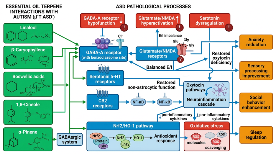
Figure 2: Molecular pathways of terpenes in autism spectrum disorder – GABA/glutamate balance, neuroinflammation, and oxidative stress
Terpenes are a diverse class of natural products that are the main components of essential oils. [B149]. Its lipophilic properties allow blood-brain barrier passage and interaction with neuronal targets. [B150]. This section analyzes molecular mechanisms of specific terpenes that target ASD-relevant pathophysiological targets.
Linalool – GABA-A/Benzodiazepine Site, Serotonin, Anti-inflammatory
Linalool (3,7-Dimethyl-1,6-octadien-3-ol) is a monoterpene alcohol and a major component of lavender (Lavandula angustifolia, 25–45 %), coriander, bergamot, and other essential oils. [B151].
GABA-A receptor modulation
Linalool shows anxiolytic effects that are blocked by flumazenil (benzodiazepine antagonist), indicating interaction with the benzodiazepine binding site of the GABA-A receptor. [B152]. In an olfactory exposure model, linalool vapors produced anxiolytic effects in mice (Elevated Plus Maze, Light-Dark Box), which were absent in anosmic mice (destruction of the olfactory epithelium), demonstrating an olfaction-mediated effect. [B152]. Electrophysiological studies show that linalool potentiates GABA-induced chloride currents in cultured neurons [B153]. This GABAergic modulation is highly relevant for ASD, as GABAergic deficits and E/I imbalance represent central pathophysiological mechanisms. [B17], [B18].
Serotonergic Modulation
Inhalation of linalool altered serotonin levels in the frontal cortex of mice, suggesting modulation of serotonergic transmission. [B154]. In a study with healthy adults, 20-minute linalool inhalation reduced depressive mood and increased parasympathetic activity (increased heart rate variability). [B155]. Ex vivo studies show that linalool increases intracellular calcium concentrations in hypothalamic oxytocin neurons, representing a potential mechanism for oxytocinergic modulation. [B155]. These findings link linalool with serotonergic and oxytocinergic systems, which are dysfunctional in ASD. [B59], [B72].
Anti-inflammatory effects
Systematic reviews report anti-inflammatory properties of linalool in preclinical models, including reduction of TNF-α, IL-6, IL-1β, and NF-κB activation. [B156]. In a stress-induced neuroinflammation model, linalool reduced microglial activation and pro-inflammatory cytokine expression. [B157]. These anti-neuroinflammatory effects are relevant for ASD, as chronic neuroinflammation is a convergent pathophysiological mechanism. [B77], [B78].
Pharmacokinetics
Following inhalation exposure, linalool is rapidly absorbed and reaches systemic circulation; metabolites (linalool oxides, glucuronides) are detected in urine. [B158]. Passage across the blood-brain barrier is likely due to lipophilicity, but direct CNS pharmacokinetic data in humans are limited. [B159].
β-Caryophyllene – CB2 Agonist, NF-κB, Neuroinflammation
Beta-caryophyllene (BCP) is a sesquiterpene hydrocarbon found in black pepper, cloves, cannabis (non-psychoactive component), copaiba, and other plants. [B160].
CB2 receptor agonism
BCP is a selective cannabinoid type 2 receptor (CB2) agonist without affinity for CB1 receptors, allowing for non-psychoactive immunomodulatory effects. [B161]. CB2 receptors are primarily expressed on immune cells (microglia, peripheral immune cells) and regulate cytokine release, microglial activation, and neuroinflammation. [B162]. In CB2 knockout mice, BCP effects were absent, which confirms CB2 specificity [B163].
Anti-neuroinflammatory effects
BCP reduces neuroinflammation in multiple preclinical models: LPS-induced neuroinflammation, traumatic brain injury, neurodegenerative diseases [B164], [B165]. Mechanisms include reduction of microglial activation (reduced Iba1, CD11b expression), suppression of pro-inflammatory cytokines (TNF-α, IL-6, IL-1β), and modulation of NF-κB signaling pathways. [B166]. In a maternal immune activation (MIA, ASD-relevant model) model, BCP treatment improved ASD-like behaviors (social deficits, repetitive behaviors) and reduced neuroinflammation in offspring. [B167].
Gabaergic and nitrinergic modulation
Behavioral pharmacological studies show that anxiolytic and antidepressant effects of BCP are blocked by flumazenil (benzodiazepine antagonist) and L-NAME (nitric oxide synthase inhibitor), indicating the involvement of GABAergic and nitrinergic systems. [B168]. These findings suggest that BCP may indirectly modulate GABAergic function via CB2-mediated neuroinflammation reduction.
Antioxidant effects
BCP shows antioxidant properties by reducing ROS, lipid peroxidation, and increasing endogenous antioxidant enzymes (SOD, catalase, GSH). [B169]. In an oxidative stress model, BCP protected neurons from H₂O₂-induced apoptosis [B170].
Clinical relevance for ASD
The combination of CB2-mediated neuroinflammation reduction, antioxidant effects, and indirect GABAergic modulation makes BCP a mechanistically plausible candidate for ASD adjunctive therapy, particularly in patients with neuroinflammatory signatures. [B171].
Boswellic acids – Nrf2/HO-1, anti-neuroinflammatory
Boswellic acids are pentacyclic triterpenic acids from the resin of Boswellia species (frankincense), including β-boswellic acid, 11-keto-β-boswellic acid (KBA), and 3-O-acetyl-11-keto-β-boswellic acid (AKBA). [B172].
Nrf2/HO-1 Activation
Boswellic acids activate the Nrf2 signaling pathway, a master regulator of the antioxidant response. [B173]. In a murine neuroinflammation model, Boswellia extract treatment increased Nrf2 nuclear localization and HO-1 expression in the hippocampus and cortex. [B174]. HO-1 catalyzes heme degradation to biliverdin (antioxidant), carbon monoxide (anti-inflammatory, vasodilatory), and iron; HO-1 induction protects against oxidative stress and neuroinflammation. [B175].
Anti-neuroinflammatory Mechanisms
Boswellic acids reduce microglial and astrocyte activation (reduced Iba1, GFAP expression), suppress pro-inflammatory cytokines (TNF-α, IL-6, IL-1β), and inhibit NF-κB and MAPK signaling pathways. [B176], [B177]. In an LPS-induced neuroinflammation model, Boswellia treatment improved cognitive deficits and reduced neuroinflammation markers. [B178].
5-Lipoxygenase (5-LOX) Inhibition
Boswellic acids, particularly AKBA, inhibit 5-LOX, an enzyme that metabolizes arachidonic acid into pro-inflammatory leukotrienes. [B179]. 5-LOX inhibition reduces leukotriene-mediated inflammation and has been associated with neuroprotective effects in preclinical studies. [B180]. However, the evidence for 5-LOX inhibition as the primary mechanism of Boswellia neuroprotection is limited in the current literature; Nrf2/HO-1 activation appears to be the dominant mechanism. [B174].
Preclinical evidence in ASD-relevant models
In a valproic acid (VPA)-induced ASD mouse model, Boswellia extract treatment improved social interaction, reduced repetitive behaviors, and normalized neuroinflammation markers. [B181]. These findings support the hypothesis that Boswellia-mediated neuroinflammation reduction and antioxidant effects can improve ASD-related behaviors.
1,8-Cineole – GABA-A/Benzodiazepine, Anxiolytic
1,8-Cineole (Eucalyptol) is a monoterpene oxide and the main component of eucalyptus (Eucalyptus globulus, 70–90 %), rosemary, tea tree, and bay. [B182].
GABA-A receptor modulation
Inhalation of 1,8-cineole produced dose-dependent anxiolytic and antidepressant effects in mice (Elevated Plus Maze, Forced Swim Test, Tail Suspension Test). [B183]. These effects were blocked by flumazenil, indicating interaction with the benzodiazepine binding site of the GABA-A receptor. [B183]. Dose dependency was demonstrated: low doses (0.1–1 µg/mouse, inhaled) showed anxiolytic effects, while higher doses (10 µg/mouse) produced sedative effects. [B184].
Anti-inflammatory and antioxidant effects
1,8-Cineole exhibits anti-inflammatory properties through inhibition of NF-κB, reduction of TNF-α, IL-1β, and leukotriene synthesis. [B185]. In an LPS-induced neuroinflammation model, 1,8-cineole reduced microglial activation and oxidative stress [B186].
Security aspects
1,8-Cineole is contraindicated in children under 3 years of age due to the risk of laryngospasm and respiratory depression. [B187]. When used correctly (inhalative, low doses), 1,8-cineole exhibits a favorable safety profile in adults and older children. [B188].
alpha-Pinene – GABAergic
alpha-Pinene is a bicyclic monoterpene found in pine trees, rosemary, sage, and cannabis. [B189].
GABAergic Modulation
Systematic reviews associate α-pinene with the modulation of GABAergic neurotransmission, although direct mechanistic studies are limited. [B190]. In behavioral models, α-pinene shows anxiolytic and sedative effects, indicating GABAergic mechanisms. [B191]. Electrophysiological data on direct GABA-A receptor interaction are lacking in the current literature.
Anti-inflammatory and neuroprotective effects
α-Pinene exhibits anti-inflammatory properties by inhibiting NF-κB and reducing pro-inflammatory cytokines [B192]. In an ischemia-reperfusion model, α-pinene protected against neuronal damage and reduced oxidative stress. [B193].
Acetylcholinesterase (AChE) Inhibition
Some studies report AChE inhibitory activity of α-pinene, which could potentiate cholinergic neurotransmission [B194]. However, relevance to ASD is unclear, as cholinergic dysfunction is not a primary pathophysiological mechanism in ASD. [B195].
Ylang-Ylang/Cananga odorata – Serotonin/Dopamine in VPA Model
Ylang-Ylang (Cananga odorata) essential oil contains linalool, geraniol, β-caryophyllene, and other terpenes. [B196].
Preclinical Evidence in the VPA-ASD Model
Inhalation of Cananga odorata essential oil improved anxiety-like behaviors, social interaction, and cognitive function in valproic acid (VPA)-induced ASD rats [B197]. Neurochemical analyses showed that Cananga odorata modulated serotonin and dopamine metabolism in the prefrontal cortex and hippocampus: increased serotonin levels, reduced 5-HIAA/serotonin ratios (indicative of reduced serotonin turnover), and normalized dopamine levels. [B197].
Mechanistic interpretation
The improvement of ASD-like behaviors by modulating serotonergic and dopaminergic systems is mechanistically plausible, as both systems are dysfunctional in ASD. [B57], [B58]. The specific components of Cananga odorata responsible for these effects have not been identified; linalool and β-caryophyllene are probable candidates based on their known mechanisms. [B152], [B161].
Limitations
These findings originate from a single preclinical model (VPA rats); replication in other ASD models and translation to human studies are lacking. [B197].
Specific Essential Oils and Clinical Evidence in ASD
Lavender/Linalool – ABA Compliance, Oxytocin Neurons
Clinical Evidence in ASD
A small pilot study (n = 12 ASD children) investigated lavender aromatherapy as an adjunct during Applied Behavior Analysis (ABA) therapy. [B198]. Lavender was diffused at “20 drops in 100 ml” (presumably water or carrier oil, details unclear) during therapy sessions. The study reported higher compliance scores (cooperation, task completion) during lavender exposure compared to control sessions. [B198]. Limitations include very small sample size, lack of randomization, unclear blinding, and lack of standardized dosing.
An observational study with four children with ASD and severe learning disabilities investigated aromatherapy massage (lavender, geranium, mandarin in carrier oil) integrated into daily routines [B199]. The study reported increased shared attention behaviors (eye contact, joint activities) after the introduction of aromatherapy massage. [B199]. Limitations include lack of control, small sample size, and confounding from massage effects (tactile stimulation, social interaction).
Mechanistic basis
The clinical findings are consistent with preclinical mechanisms: linalool-mediated GABAergic modulation can reduce anxiety and improve cooperation; serotonergic and oxytocinergic modulation can promote social attention and interaction. [B152], [B155]. A study with healthy adults showed that lavender inhalation reduced depressive mood and, in ex vivo experiments, increased intracellular calcium concentrations in hypothalamic oxytocin neurons. [B155]. This oxytocinergic modulation is highly relevant for ASD, as oxytocin dysfunction is an established pathophysiological mechanism [B72].
Limitations of the Evidence
The clinical evidence for lavender in ASD is of very low quality: small sample sizes, lack of randomization, insufficient blinding, heterogeneous interventions, and lack of replication. [B198], [B199]. High-quality, randomized controlled trials with standardized lavender preparations, objective outcome measures, and sufficient power are lacking.
Bergamot – RCT in children with ASD, outcome not significant
Randomized controlled trial
A randomized, blinded study investigated bergamot (Citrus bergamia) essential oil for medically-induced anxiety in children with ASD (n=42). [B200]. Children were randomized to 15-minute inhalation of bergamot oil or placebo (water) before medical examinations. The primary outcome was anxiety measured with the Modified Yale Preoperative Anxiety Scale (mYPAS). After adjustment for baseline anxiety, the bergamot group showed no significant reduction in anxiety compared to placebo (p > 0.05). [B200].
interpretation
This is the only identified randomized controlled trial of essential oils in children with ASD. The negative outcome could have several causes: (1) short exposure duration (15 minutes) may be insufficient; (2) acute anxiety in medical settings may be harder to modulate than chronic anxiety; (3) individual variability in olfactory sensitivity and preferences; (4) possible ceiling effects with already high baseline anxiety. [B200].
Preclinical mechanisms
Preclinical studies show anxiolytic effects of bergamot in rodent models [B201]. Interestingly, these effects were not blocked by flumazenil, suggesting non-benzodiazepine-mediated mechanisms and distinguishing bergamot from linalool and 1,8-cineole. [B201]. Bergamot contains linalool, linalyl acetate, limonene, and bergapten; the specific components responsible for anxiolytic effects are unclear. [B202].
Clinical implications
The negative result of this RCT dampens expectations for bergamot in acute anxiety in ASD. Further studies with longer exposure durations, chronic anxiety outcomes, and different ASD populations are needed before bergamot can be recommended for ASD. [B200].
Frankincense / Boswellia – Anti-neuroinflammatory, Nrf2
Clinical Evidence in ASD
No randomized controlled trials with frankincense (Boswellia) essential oil or Boswellia extracts in ASD patients were identified. The present literature does not include ASD-specific clinical data for frankincense. [B203].
Preclinical evidence
As described in Section 3.3, boswellic acids show robust anti-neuroinflammatory and antioxidant effects in preclinical models, including Nrf2/HO-1 activation, microglia inhibition, and improvement of ASD-like behaviors in VPA mouse models [B174], [B181]. This mechanistic plausibility supports the hypothesis that Boswellia may be useful in ASD patients with neuroinflammation signatures.
Clinical application of other indications
Boswellia extracts (oral) are used clinically for inflammatory diseases (arthritis, asthma, inflammatory bowel diseases), with moderate evidence of efficacy and a favorable safety profile. [B204]. Inhalant use of frankincense essential oil is traditional, but clinical pharmacokinetic and efficacy data are lacking [B205].
Research needs
Controlled clinical trials with Boswellia (oral or inhaled) in ASD patients, ideally stratified by neuroinflammation biomarkers, are needed to evaluate clinical efficacy. [B206].
Cedarwood – Sedation, GABAergic
Clinical and preclinical evidence
The present literature contains no specific mechanistic or clinical data for cedarwood (Cedrus atlantica, Juniperus virginiana) essential oil in ASD or in ASD-relevant preclinical models. [B207]. Traditional applications include sedation and anxiety reduction, but scientific evidence for GABAergic mechanisms or clinical efficacy is lacking in the analyzed literature [B208].
Main components
Cedarwood oil contains cedrol (sesquiterpene alcohol), α-cedrene, and thujopsene. [B209]. Cedrol is associated with sedative properties, but direct studies on GABA-A receptor interaction or neuroinflammation modulation are lacking [B210].
Conclusion
Evidence for cedarwood in ASD is insufficient; recommendations cannot be made based on the existing literature. [B207].
Vetiver – Attention
Clinical and preclinical evidence
The present literature contains no specific data for vetiver (Vetiveria zizanioides) essential oil in ASD or in ASD-relevant models [B211]. Anecdotal reports and traditional uses suggest effects on attention and hyperactivity, but controlled studies are lacking. [B212].
Main components
Vetiver oil contains vetiverol, khusimol, alpha-vetivone, and beta-vetivone (sesquiterpene alcohols and ketones). [B213]. Mechanistic studies of these components with regard to neurotransmitter systems or neuroinflammation are lacking in the analyzed literature. [B214].
Conclusion
Evidence for vetiver in ASD is insufficient; further research is needed before recommendations can be made. [B211].
Ylang-Ylang – Serotonin/Dopamine, VPA Model
Preclinical evidence
As described in Section 3.5, inhalation of Cananga odorata (ylang-ylang) essential oil improved ASD-like behaviors in VPA-rats and modulated serotonin and dopamine metabolism in the prefrontal cortex and hippocampus. [B197]. This is the only identified study using Ylang-Ylang in an ASD-relevant model.
Clinical Evidence in ASD
No clinical studies with ylang-ylang in ASD patients were identified. [B215].
Clinical application of other indications
Ylang-ylang is traditionally used for anxiety, mood, and relaxation. A small study in healthy adults showed that ylang-ylang inhalation reduced blood pressure and heart rate, and increased subjective calmness. [B216]. Mechanistic studies on serotonergic and dopaminergic effects in humans are lacking [B217].
Conclusion
The preclinical evidence is promising, but translation to human ASD studies is lacking. Ylang-ylang could be considered as a candidate for clinical trials in ASD, particularly for anxiety and mood symptoms. [B197].
Further indicated essential oils for ASD
Based on a current literature review, the following additional essential oils were identified that may be relevant for ASD through neurobiologically plausible mechanisms.
Ginger (Zingiber officinale) – AKT/GSK3β Signaling Pathway
Active ingredients
6-Gingerol
6-Shogaol
Zingiberene
beta-Sesquiphellandrene
Receptors and Mechanisms of Action
AKT/GSK3β signaling pathway: Increased phosphorylation → improved synaptic plasticity
– Anti-neuroinflammatory: Reduction of STAT3 phosphorylation and TNF-α
– Neurogenesis: Improved hippocampal neurogenesis markers in preclinical models
Glio-inhibition: Reduced reactive astrogliosis
Clinical/Preclinical Evidence
- Preclinical study (VPA mouse model)
Oral ginger extract (4 weeks, starting from the 6th week of life) significantly improved social interaction, reduced anxiety-like behavior, and enhanced memory performance. [B347]
– Mechanistic: Upregulation of AKT/GSK3β, inhibition of STAT3/TNF-α, improved synapse formation
– Clinical RCTs in ASD are still lacking, but preclinical evidence is strong and mechanistically well-founded
Dosage
2–3 drops for room 15 to 25 m²
– Combination with lavender (1:2) for a relaxing effect
– Application: 30 min before social activities
Security
Well tolerated; use with caution in children < 3 years old.
Sandalwood (Santalum album) – α/β-Santalol
Active ingredients
α-Santalol (50–55%)
β-Santalol (20–25%)
Sesquiterpene
Receptors and Mechanisms of Action
– Serotonergic Modulation: α-Santalol modulates 5-HT receptors (preclinical)
– Anxiolytic: GABAergic activity documented in animal models
– Sedating/Calming: Suitable for sensory hypersensitivity in ASD
Clinical evidence
– Sandalwood was part of the Hiwa Syrup RCT (Persian medicine, multiple herbs) in ASD children, effects not isolatable [B348]
– Individual studies on anxiolysis in healthy subjects show a calming effect
– Direct ASD-specific studies with pure sandalwood oil are lacking
Dosage
- 3-4 drops for rooms 15 to 25 m²
– Especially suitable for evening/sleep rituals for children with ASD who have trouble falling asleep
– Recommended combination with lavender (1:1)
Security
Very well tolerated; one of the safest essential oils for children (from 2 years of age).
Terpene Blends (Cannabis-Inspired) – Prosocial Effects
Active ingredients
Myrcene
Limes
Linalool
Pine
β-Caryophyllene (as a blend)
Receptors and Mechanisms of Action
– CB1/CB2 Agonism: Terpene mixtures enhance CBD effects (Entourage effect)
GABAergic Modulation: Linalool + Myrcene synergistically
– Prosocial effect demonstrated regardless of CBD
Clinical evidence
- BTBR mouse model (ASD model)
Inhaled terpene blends (myrcene, limonene, linalool, pinene, β-caryophyllene) showed acute prosocial effects independently of CBD [B349] – Terpene mixtures enhanced CBD effect with combined application
– Hint at entourage effect as a therapeutic principle for ASD
Dosage
– Blend Recommendation: Lavender (2 tsp) + Black Pepper (1 tsp) + Bergamot (1 tsp) + Rosemary (1 tsp)
– For Room 15, it's 25 m²: 5 drops total mixture
– Application: 30 min before social activities or group therapy
Dosage recommendations for Organik Aromas Nebulizer 3.0
The Organik Aromas Nebulizer 3.0 uses the Venturi principle (Bernoulli effect) for waterless cold nebulization of essential oils. [B218]. Unlike ultrasonic or heat diffusers, which dilute oils with water or heat them, the nebulizer produces fine aerosol particles (1 to 5 µm) of pure essential oils without thermal degradation. [B219]. This allows for higher concentrations of volatile terpenes in the room air and potentially stronger pharmacological effects. [B220].
Venturi principle and cold fogging
The Venturi principle describes the reduction in pressure within a fluid as it flows through a constriction. [B221]. In the nebulizer, compressed air is directed through a nozzle, which creates a negative pressure that draws essential oil from a reservoir and atomizes it into fine droplets. [B222]. These droplets are released into the air, where they are inhaled or settle on surfaces. [B223].
Benefits of cold fogging
– Receipt of thermolabile components (no heat degradation) [B224]
- Higher concentrations of volatile terpenes (no water dilution effect) [B225]
Fine particle size (1 to 5 µm) enables deep airway penetration [B226]
Disadvantages
- Higher costs compared to ultrasonic diffusers [B227]
– Increased oil consumption [B228]
Potential for overdose with improper use [B229]
Room size-specific drizzle amounts
Dosage recommendations are based on room volume, air exchange rate, desired terpene concentration, and exposure duration. The following recommendations are conservative and consider ASD-specific sensory hypersensitivity. [B230].
Table 1: Room size-specific dosage recommendations for Organik Aromas Nebulizer 3.0
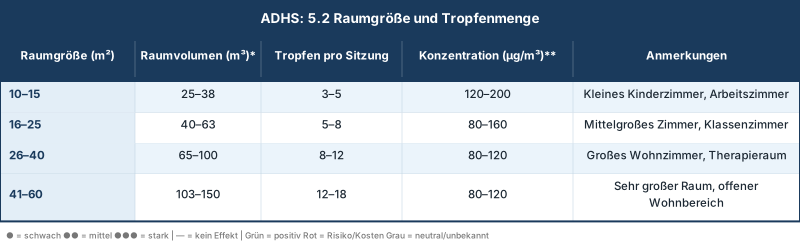
Notes
- Drop volume 1 drop ≈ 0.05 ml essential oil [B231]
- Starting dose: Start with the lowest recommended drop count and gradually increase over 1 to 2 weeks to assess tolerance. [B232]
- Customization ASD patients with pronounced olfactory hypersensitivity may require lower doses or longer breaks [B233]
- Ventilation: Good room ventilation is essential to avoid excessive accumulation [B234]
Application duration and breaks
Application duration
15 to 30 minutes per session are sufficient to achieve therapeutic terpene concentrations based on preclinical inhalation studies. [B183], [B184]. Longer applications (> 30 minutes) increase the risk of sensory overload and olfactory adaptation (reduced perception with continuous exposure). [B235].
Breaks
30 to 60-minute breaks between applications allow for olfactory recovery, metabolism of inhaled terpenes, and reduction of indoor air concentrations. [B236]. Continuous exposure without breaks can lead to headaches, nausea, and sensory aversion, especially in ASD patients with sensory hypersensitivity. [B237].
Daily Applications
2 to 3 applications per day (morning, afternoon, evening) are sufficient for most indications [B238]. More than 4 applications per day increase the risk of side effects without additional therapeutic benefit [B239].
Time of day specific protocols
Morning (activating, focusing)
- Oils Rosemary (1,8-cineole, α-pinene), Peppermint (menthol, menthone), Lemon (limonene), Bergamot (linalool, linalyl acetate)
- Dosage: 3-5 drops for 20-30 m² room
- Duration: 15-20 minutes
- Target: Promoting alertness, attention, and cognitive function for school/therapy [B240]
Afternoon (balancing, mood-modulating)
- Oils Bergamot, Ylang-Ylang, Geranium, Frankincense
- Dosage: 3-5 drops for 20-30 m² room
- Duration: 20-30 minutes
- Target: Reduction of afternoon anxiety, mood stabilization, transition between activities [B241]
Evening (calming, sleep-inducing)
- Oils Lavender (Linalool, Linalyl acetate), Roman Chamomile (Esters), Cedarwood (Cedrol), Vetiver
- Dosage: 4–6 drops for 20–30 m² room
- Duration: 30 minutes before bedtime
- Target: Reduction of evening anxiety, promoting relaxation, and sleep initiation [B242]
ASD-specific characteristics
Sensory hypersensitivity 60-90 % of ASD patients show olfactory hypersensitivity or aversion to certain smells [B243]. Graduated introduction is essential:
- Week 1: Exhibition outside the room (oil diffusing into adjacent room) to gauge acceptance [B244]
- Week 2: Lowest dose (2 drops) for 10 minutes, patient outside the room during nebulization. [B245]
- Week 3: Lowest dose, patient in room, observation for signs of tolerance (relaxation, no aversion) [B246]
- Week 4+ Graduated increase in dose and duration based on individual tolerance [B247]
Preference-based selection
Have the patient smell various oils (if possible) and express preferences. Olfactory preference correlates with therapeutic acceptance and compliance. [B248].
Visual and auditory cues
Use visual timers or auditory cues to indicate the start and end of aromatherapy sessions, increasing predictability and control for ASD patients [B249].
Parent/Caregiver Training
Train parents and caregivers to observe signs of tolerance (relaxation, improved attention) and aversion (grimacing, withdrawal, agitation). [B250].
Quality Requirements – GC/MS Analysis
Gas Chromatography-Mass Spectrometry (GC/MS)
GC/MS is the gold standard for chemical characterization of essential oils [B251]. GC separates volatile components based on boiling point and polarity; MS identifies components based on mass-to-charge ratio. [B252]. GC/MS analysis allows:
- Identification of main components: Confirmation of the presence of therapeutically relevant terpenes (e.g., linalool in lavender, 1,8-cineole in eucalyptus) [B253]
- Quantification Percentage composition of each component [B254]
- Forgery detection Identification of synthetic additives, diluents, or contaminants [B255]
- Batch-to-Batch Consistency Ensuring reproducible composition [B256]
Quality Criteria for Therapeutic Essential Oils
- 100 % rein
No synthetic additives, carrier oils, or diluents [B257] - Therapeutic grade
Oils from plants that were grown, harvested, and distilled under optimal conditions B258 - Organisch/Wildsammlung
Free from pesticides, herbicides, and chemical fertilizers [B259] - GC/MS Certificate
Each batch should be supplied with a GC/MS analysis certificate documenting the main components and purity. B260 - Botanical name
Unique identification of the plant species (e.g., Lavandula angustifolia, not just “Lavender”) [B261] - Country of origin and distillation date
Transparency about origin and freshness [B262]
Storage
Essential oils should be stored in dark glass bottles at a cool temperature (<77°F) to minimize oxidation and degradation. [B263]. Oxidized oils can cause skin irritation and show reduced therapeutic effectiveness. [B264].
Safety Instructions and Contraindications
ASD-specific sensory hypersensitivity
Sensory processing disorders are present in 60–90 % of ASD patients and manifest as hypersensitivity or hyposensitivity to sensory stimuli. [B265]. Olfactory hypersensitivity can lead to aversion, anxiety, nausea, and avoidance behavior. [B266].
Risk Management
- Graduate Introduction As described in Section 5.4, begin with minimal doses outside the patient room [B244]
- Preference Screening Test oils for therapeutic use to identify adverse reactions [B248]
- Demolition criteria: Stop immediately at any signs of distress (agitation, withdrawal, nausea, headache) [B267]
- Alternative Application Methods: For inhalational intolerance, consider topical application (diluted in carrier oil, patch test required) or passive diffusion (oil on a cotton pad in a room corner). [B268]
Epilepsy Risk - Camphor-Containing Oils
10–30 % of ASD patients have comorbid epilepsy [B269]. Certain essential oils, particularly those containing camphor, can lower seizure thresholds and trigger seizures. [B270].
Contraindicated oils for epilepsy
- Camphor (Cinnamomum camphora): Contains camphor (up to 50 %), a known convulsant [B271]
- Rosemary (Rosmarinus officinalis, Camphor chemotype): Contains 15–25 % camphor [B272]
- Sage (Salvia officinalis): Contains thujone (10–40 %), a GABA-A antagonist with convulsive properties [B273]
- Hyssop (Hyssopus officinalis): Contains pinocamphone and isopinocamphone, which can trigger seizures [B274]
- Fennel (Foeniculum vulgare): Contains trans-anethole, which can have convulsant effects in high doses [B275]
Safe Alternatives in Epilepsy: – Lavender (Lavandula angustifolia, Linalool chemotype): Anticonvulsant properties in preclinical studies [B276] – Roman Chamomile (Anthemis nobilis): No known proconvulsant effects [B277] – Bergamot (Citrus bergamia, bergapten-free): Safe for epilepsy [B278]
Clinical Recommendation: For ASD patients with epilepsy, only oils without camphor or thujone components should be used. Consultation with a neurologist before introducing aromatherapy is advisable. [B279].
Eucalyptus in children under 3 years old
1,8-Cineole (the main component of eucalyptus) is contraindicated in children under 3 years of age due to the risk of laryngospasm, bronchospasm, and respiratory depression. [B280]. Mechanisms include direct irritation of the airway mucosa and triggering of vagal reflexes. [B281].
Contraindicated oils in children <3 years old
– Eucalyptus (Eucalyptus globulus, E. radiata): 70–90 % 1.8-Cineole [B282]
– Rosemary (Rosmarinus officinalis, 1,8-Cineol chemotype): 40–50 % 1,8-Cineol [B283]
– Tea Tree (Melaleuca alternifolia): 5–15 % 1,8-cineole (lower risk, but caution advised) [B284]
Peppermint (Mentha piperita): Menthol can cause similar respiratory irritation [B285]
Safe alternatives for children under 3 years old
– Lavender (Lavandula angustifolia): Safe from birth with proper dilution [B286]
Roman Chamomile (Anthemis nobilis): Safe from birth [B287]
– Mandarin (Citrus reticulata): Safe from 6 months [B288]
Clinical recommendation
Avoid oils rich in 1,8-cineole in children with ASD under 3 years of age.
For children 3–6 years old, use low doses (50 % of the adult dose) and monitor carefully for respiratory symptoms. [B289].
Interactions with antipsychotics: Risperidone, Aripiprazole
Risperidone and aripiprazole are the only FDA-approved medications for irritability in ASD. [B290]. Both are metabolized by cytochrome P450 enzymes (CYP2D6, CYP3A4). [B291]. Some essential oils and terpenes can inhibit or induce CYP enzymes, which can lead to altered drug levels [B292].
Potential interactions:
Bergamot
Contains bergapten (furanocoumarin), which inhibits CYP3A4 [B293]. Theoretically, bergamot could increase risperidone and aripiprazole levels, which could intensify side effects (sedation, extrapyramidal symptoms). [B294]. However, inhalation exposure is significantly lower than oral intake (grapefruit juice effect); the clinical relevance of inhaled bergamot exposure is unclear. [B295].
Recommendation
Use bergapten-free bergamot (FCF, furanocoumarin-free) in patients on risperidone/aripiprazole [B296].
Grapefruit
Grapefruit essential oil contains furanocoumarins and inhibits CYP3A4 [B297]. When patients are taking CYP3A4-metabolized medications [B298].
St. John's wort (Hypericum perforatum)
Although not a typical essential oil, St. John's Wort is sometimes mentioned in aromatherapy contexts. St. John's Wort is a potent CYP3A4 inducer and can reduce risperidone/aripiprazole levels, leading to a loss of efficacy. [B299]
Contraindication Avoid St. John's Wort in patients taking antipsychotics [B300].
Lavender, Chamomile, Cedarwood
No known clinically relevant CYP interactions [B301]. Safe in patients on risperidone/aripiprazole [B302].
Pharmacodynamic Interactions
Sedative essential oils (lavender, chamomile, cedarwood) can have additive sedative effects with antipsychotics [B303]. This can be therapeutically desirable (e.g., promoting sleep), but requires monitoring for excessive sedation. [B304].
Clinical recommendation
Inform prescribing physicians about aromatherapy use. Use bergapten-free oils in patients under CYP3A4 substrates. Monitor for altered drug effects (sedation, side effects) [B305].
Quality Requirements: GC/MS Analysis
As described in Section 5.5, GC/MS analysis is essential to ensure purity, composition, and the absence of contaminants. [B251]. Adulterated or contaminated oils can cause allergies, skin irritation, respiratory symptoms, and unpredictable pharmacological effects. [B306].
Common Alterations
- Synthetic Additives Synthetic Linalool or Linalyl Acetate in “Lavender” Oil [B307]
- Thinner Propylene glycol, dipropylene glycol methyl ether (DPM) [B308]
- Cheaper oils: Lavandin (Lavandula x intermedia) sold as true lavender (Lavandula angustifolia) [B309]
- Pesticides: Residues of pesticides, herbicides in non-organic oils [B310]
Security recommendation
Only use GC/MS certified, 100 % pure, organic essential oils from trusted suppliers. Request GC/MS certificates for each batch [B311].
discussion
Evidence
The evidence for essential oils in ASD is heterogeneous and predominantly of low quality. The clinical literature includes one negative randomized controlled trial (bergamot for acute anxiety). [B200], small pilot studies with methodological limitations (lavender in ABA compliance) [B198], Observational studies (aromatherapy massage) [B199] and case reports [B312]. No study demonstrates efficacy for core ASD symptoms (social communication, repetitive behaviors) with sufficient evidence quality. [B313].
In contrast, the preclinical evidence is robust: multiple studies demonstrate that specific terpenes (linalool, β-caryophyllene, boswellic acids, 1,8-cineole) target ASD-relevant molecular targets (GABA-A receptors, CB2 receptors, Nrf2 pathway, neuroinflammation) and improve ASD-like behaviors in animal models. [B152], [B161], [B174], [B183], [B197]. This discrepancy between preclinical plausibility and clinical evidence is typical for complementary therapies and reflects a lack of research funding, methodological challenges, and translation gaps. [B314].
Mechanistic plausibility
The mechanistic plausibility for essential oils in ASD is given, based on convergence between ASD pathophysiology and terpene mechanisms of action:
GABA/Glutamate E/I Imbalance
Linalool and 1,8-cineole modulate GABA-A receptors at the benzodiazepine binding site, which could potentiate inhibitory neurotransmission and normalize E/I balance. [B152], [B183]. These mechanisms are analogous to bumetanide, which showed moderate improvements in ASD in clinical studies [B52].
Neuroinflammation
Beta-caryophyllene (CB2 agonist) and boswellic acids (Nrf2 activator) reduce microglial activation, pro-inflammatory cytokines, and oxidative stress in preclinical models [B161], [B174]. As neuroinflammation is an established pathophysiological mechanism in ASD [B77], could these terpenes be useful in patients with neuroinflammation signatures (elevated peripheral cytokines, PET evidence of microglial activation) [B315].
Serotonin/Dopamine
Linalool and Ylang-Ylang modulate serotonergic and dopaminergic systems in preclinical studies [B154], [B197]. These mechanisms could improve comorbid symptoms (anxiety, mood), though effects on core symptoms are unlikely. [B316].
Oxytocin
Linalool increases intracellular calcium concentrations in oxytocin neurons [B155]. Oxytocin regulates GABAergic development and improved ASD-like phenotypes in preclinical studies. [B50], could linalool indirectly modulate ox tonergic signaling pathways [B317].
Gut-Brain Axis
Although direct evidence is lacking, anti-inflammatory terpenes (β-caryophyllene, boswellic acids) could reduce systemic inflammation and indirectly modulate gut-brain axis pathways. [B318]. This hypothesis requires experimental validation. [B319].
Limitations
Small studies
Most ASD-specific studies have sample sizes <20, meaning insufficient power to detect moderate effects. [B320]. Meta-analyses are not possible due to heterogeneity. [B321].
No RCTs for core disorders
There are no randomized controlled trials examining the efficacy of essential oils for core ASD symptoms (social communication, repetitive behaviors). [B322]. The only identified RCT (bergamot) was negative for acute anxiety. [B200].
Methodological heterogeneity
Studies vary in oil selection, dosage, route of administration (inhalation, massage, topical), exposure duration, outcome measures, and populations [B323]. This heterogeneity prevents synthesis and comparability [B324].
Missing biomarkers
No study stratifies patients by neurobiological subtypes (e.g., neuroinflammation signatures, E/I imbalance markers) or uses objective biomarkers as outcomes. [B325]. Precision medicine approaches could identify responders. [B326].
Publication bias
Positive case reports and small pilot studies are more likely to be published than negative studies, which can lead to an overestimation of effects. [B327].
Translation Gap
Preclinical models (VPA rats, MIA mice) do not replicate the full complexity of human ASD heterogeneity [B328]. Terpene doses in animal studies are often higher than what is achievable with human inhalation exposure [B329].
Placebo effects
Aromatherapy is associated with strong expectation effects; blinding is difficult due to obvious smells [B330]. The bergamot RCT used water as a placebo, which means insufficient blinding. [B200].
Safety Data Long-term safety data for chronic inhalational exposure in children are lacking [B331]. Most safety data originates from topical or oral application [B332].
Outlook
Research priorities
- High-quality RCTs Randomized, double-blind, placebo-controlled trials with sufficient power (n > 100), standardized oil preparations (GC/MS certified), objective outcome measures (biomarkers, actigraphy, eye-tracking), and longer intervention durations (8–12 weeks) [B333].
- Stratification by subtype: Identification of responders based on neurobiological subtypes (neuroinflammation markers, E/I imbalance, microbiome profiles) [B334]. Precision medicine approaches could increase effect sizes B335.
- Mechanistic Studies Human studies with biomarker outcomes (cytokine profiles, neuroimaging, EEG, microbiome analyses) before and after intervention to validate mechanisms [B336].
- Pharmacokinetics Human Pharmacokinetic Studies for Inhalative Terpene Exposure: Plasma Concentrations, CNS Penetration, Metabolism, Dose-Response Relationships [B337].
- Combination Therapies: Investigation of Synergies between Essential Oils and Established Interventions (Behavioral Therapy, Pharmacotherapy, Microbiome Interventions) [B338].
- Long-term safety Prospective Cohort Studies on the Long-Term Safety of Chronic Inhalative Exposure in Children [B339].
- Personalized Aromatherapy: Development of algorithms for oil selection based on individual symptom profiles, preferences, and neurobiological markers. [B340].
Clinical implications
Based on current evidence, essential oils can be considered as part of a multimodal therapeutic approach for comorbid symptoms (anxiety, sleep disorders, sensory dysregulation) in ASD, but not as monotherapy or a substitute for evidence-based core interventions. [B341]. Clinicians should:
- Communicating Realistic Expectations: Essential Oils Can Alleviate Comorbid Symptoms but Cannot Cure Core Symptoms [B342]
- Consider individual tolerance and preferences [B343]
- High-quality, GC/MS-certified oils [B344]
- Consider safety aspects (epilepsy, age, medication interactions) [B345]
- Monitor effects and adjust therapy [B346]
Conclusion
Essential oils represent a mechanistically plausible, but clinically insufficiently validated, adjunctive therapy option for autism spectrum disorder. The neurobiological heterogeneity of ASD – characterized by GABA/glutamate E/I imbalance, neuroinflammation, oxidative stress, gut-brain axis alterations, and synaptic dysfunction – offers multiple therapeutic targets that specific terpenes can address.
Preclinical evidence demonstrates that linalool (GABA-A modulation, serotonergic regulation), β-caryophyllene (CB2-mediated neuroinflammation reduction), boswellic acids (Nrf2/HO-1 activation), 1,8-cineole (anxiolytic GABAergic effects), and ylang-ylang (serotonin/dopamine modulation) modulate ASD-relevant pathophysiological mechanisms and improve ASD-like behaviors in animal models. This mechanistic plausibility is supported by traditional uses and limited human data for anxiety, sleep, and mood.
However, the clinical evidence in ASD is of low quality: a negative RCT (bergamot), small pilot studies with methodological limitations (lavender and ABA compliance), and observational studies. No study demonstrates efficacy for core ASD symptoms with sufficient evidence quality. High-quality, randomized controlled trials with standardized preparations, objective outcomes, and adequate power are lacking.
For the Organik Aromas Nebulizer 3.0, room size-specific dosage recommendations are being developed (2–10 drops for 10–60 m², 15–30 minutes, 30–60 minute breaks), with time-of-day adjusted protocols (activating in the morning, calming in the evening) and ASD-specific considerations (graduated introduction, preference screening, sensory hypersensitivity). Safety aspects include contraindications for epilepsy (camphor-containing oils), children < 3 years old (eucalyptus), potential interactions with antipsychotics (bergapten-containing oils), and the need for GC/MS-certified, pure oils.
Essential oils can be considered as part of a multimodal therapeutic approach for comorbid symptoms (anxiety, sleep disturbances, sensory dysregulation) in ASD, but they do not replace evidence-based core interventions (behavioral therapy, structured education). Future research should prioritize high-quality RCTs, stratification by neurobiological subtypes, mechanistic biomarker studies, and long-term safety data to validate the therapeutic potential of essential oils in ASD and develop precision medicine approaches.
Joint conclusion
Overarching Insights: ADHD and Autism
Both neurodevelopmental disorders, ADHD and Autism Spectrum Disorder (ASD), share fundamental neurobiological commonalities, making adjunctive therapy with essential oils plausible for both indications.
Common neurobiological target structures:
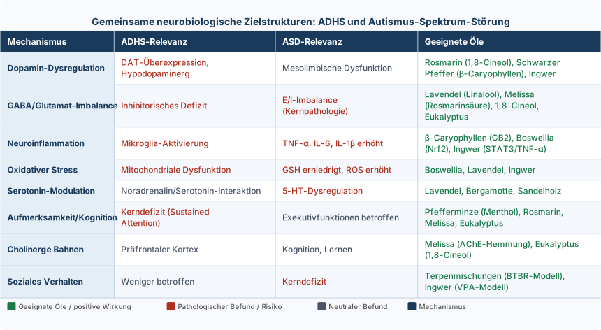
Joint application recommendation (Organik Aromas Nebulizer 3.0):
For both indications, the Venturi cold nebulization principle without water or heat is the optimal application method: – No thermal degradation of terpenes – Precise particle size for olfactory absorption (< 5 µm) – GC/MS-pure oils retain their molecular integrity
Important Notice
Adjuvant therapy with essential oils does not replace evidence-based standard therapy (methylphenidate for ADHD, ABA/behavioral therapy for ASD). It is a complementary addition that should be individualized and carried out under expert guidance. The evidence for both indications is still limited; larger RCTs are needed.
References ADHD
Chen et al. (2008). The effect of applying cinnamon aromatherapy for children with attention deficit hyperactivity disorder.https://pmc.ncbi.nlm.nih.gov/articles/PMC8538900/
Chen et al. The effect of applying cinnamon aromatherapy for children with attention deficit hyperactivity disorder.https://pmc.ncbi.nlm.nih.gov/articles/PMC8538900/
[A3] Godfrey (2001). The role of essential oils in the treatment and management of attention deficit hyperactive disorder. International Journal of Aromatherapy.https://doi.org/10.1016/S0962-4562(01 )80035-7
[A4] Godfrey. Essential oils: Complementary treatment for attention deficit hyperactive disorder (ADHD).https://www.researchgate.net/publication/248871372_The_role_of_essential_oils_in_the_treatment_and_management_of_attention_deficit_hyperactive_disorder
[A5] Godfrey. The Role of Essential Oils in the Treatment and Management of Attention Deficit Hyperactive Disorder (ADHD).https://www.researchgate.net/publication/248871372_The_role_of_essential_oils_in_the_treatment_and_management_of_attention_deficit_hyperactive_disorder
[A6] Ding et al. (1994). Pharmacokinetics and in vivo specificity of [LLC]dl-threo-methylphenidate for the presynaptic dopaminergic neuron. Synapse.https://doi.org/10.1002/SYN.890180207
[A7] Volkow et al. (2002). Mechanism of action of methylphenidate: insights from PET imaging studies. Journal of Attention Disorders.https://doi.org/10.1177/070674370200601S05
[A8] Silva (2024). Aromatherapy in the Care of an Atypical Child with ADHD and Atopic Dermatitis.https://doi.org/10.62435/2965-7253.bjhae.2024.iisinaroma1
[A9] Volkow et al. (1998). Dopamine Transporter Occupancies in the Human Brain Induced by Therapeutic Doses of Oral Methylphenidate. American Journal of Psychiatry.https://doi.org/10.1176/AJP.155.10.1325
[A10] Jacob et al. (2024). Use of aromatherapy in student support.https://doi.org/10.37885/240717094
[A11] Dipasquale et al. (2020). Unravelling the effects of methylphenidate on the dopaminergic and noradrenergic functional circuits. bioRxiv.https://doi.org/10.1101/2020.03.09.983528
[A12] Volkow et al. (2002). Role of dopamine in the therapeutic and reinforcing effects of methylphenidate in humans: results from imaging studies. European Neuropsychopharmacology.https://doi.org/10.1016/S0924-977X(02 )00104-9
[A13] Spencer et al. (2006). PET Study Examining Pharmacokinetics, Detection and Likeability, and Dopamine Transporter Receptor Occupancy of Short- and Long-Acting Oral Methylphenidate. American Journal of Psychiatry.https://doi.org/10.1176/APPI.AJP.163.3.387
Castillo et al. (2018). Essential oils as modifiers of human behavior. Tropical and Subtropical Agroecosystems.https://www.revista.ccba.uady.mx/ojs/index.php/TSA/article/view/2583
Crunelle et al. (2014). Dopamine transporter occupancy by methylphenidate and impulsivity in adult ADHD. British Journal of Psychiatry.https://doi.org/10.1192/BJP.BP.113.132977
[A16] Ding et al. (1997). Chiral drugs: comparison of the pharmacokinetics of [11C]d-threo and l-threo-methylphenidate in the human and baboon brain. Psychopharmacology.https://doi.org/10.1007/S002130050267
[A17] Spencer et al. (2009). A PET study examining pharmacokinetics and dopamine transporter occupancy of two long-acting formulations of methylphenidate in adults. International Journal of Molecular Medicine.https://doi.org/10.3892/IJMM_00000339
[A18] Marcel et al. (2024). Responsivity of the Striatal Dopamine System to Methylphenidate—A Within-Subject I-123-β-CIT-SPECT Study in Male Children and Adolescents With Attention-Deficit/Hyperactivity Disorder.https://doi.org/10.5283/epub.57629
Kennedy et al. (2018). Volatile Terpenes and Brain Function: Investigation of the Cognitive and Mood Effects of Mentha × Piperita L. Essential Oil with In Vitro Properties Relevant to Central Nervous System Function. Nutrients.https://doi.org/10.3390/NU10081029
[A20] Batiha et al. (2023). A review of the bioactive components and pharmacological properties of Lavandula species. Naunyn-Schmiedeberg's Archives of Pharmacology.https://doi.org/10.1007/s00210-023-02392-x
[A21] Guzmán-Gutiérrez et al. (2015). Linalool and β-pinene exert their antidepressant-like activity through the monoaminergic pathway.https://pubmed.ncbi.nlm.nih.gov/25771248/
[A22] Martinez et al. (2020). Ventral striatum supports methylphenidate's therapeutic effects on impulsive choices expressed in a temporal discounting task. Scientific Reports.https://doi.org/10.1038/s41598-020-57595-6
[A23] López et al. (2017). Exploring Pharmacological Mechanisms of Lavender (Lavandula angustifolia) Essential Oil on Central Nervous System Targets. Frontiers in Pharmacology.https://doi.org/10.3389/FPHAR.2017.00280
[A24] Rathnayake (2025). “Neuroaromatics”: Aromatherapy and the Limbic Stress Response.https://doi.org/10.4038/jssnisd.v3i1.28
[A25] Aster et al. (2021). Responsivity of the striatal dopamine system to methylphenidate – a within-subject I-123-β-CIT-SPECT study in children and adolescents with Attention-Deficit/Hyperactivity Disorder. medRxiv.https://doi.org/10.1101/2021.11.19.21265993
Cardoso et al. (2025). The Impact of Lavender Essential Oil on the Human Nervous System: A Review. Deleted Journal.https://doi.org/10.62435/2965-7253.bjhae.2025.bjhae22
[A27] Moss et al. (2003). Aromas of rosemary and lavender essential oils differentially affect cognition and mood in healthy adults. International Journal of Neuroscience.https://doi.org/10.1080/00207450390161903
[A28] Sinha et al. (2005). Complementary and alternative medicine use in children with attention deficit hyperactivity disorder. Journal of Paediatrics and Child Health.https://doi.org/10.1111/J.1440-1754.2005.00530.X
[A29] Springer et al. (2018). ADHD: The Lifespan and Effects from Adolescence through Adulthood.https://dergipark.org.tr/en/pub/pgy/548052.xml
Colzato et al. (2014). Attentional control in the attentional blink is modulated by odor. Attention Perception & Psychophysics.https://doi.org/10.3758/S13414-014-0733-0
Kennedy, D. O., Scholey, A., Tildesley, N. T. J., Perry, E. K., & Wesnes, K. A. (2002). Modulation of mood and cognitive performance following acute administration of Melissa officinalis (lemon balm). Pharmacology, Biochemistry, and Behavior, 72(4), 953–964. https://doi.org/10.1016/S0091-3057(02)00777-3
A32. Müller, S. F., & Klement, S. (2006). A combination of valerian and lemon balm is effective in the treatment of restlessness and dyssomnia in children. Phytomedicine, 13(6), 383–387. https://doi.org/10.1016/j.phymed.2006.01.013
Scholey, A., Gibbs, A., Neale, C., et al. (2014). Anti-stress effects of lemon balm-containing foods. Nutrients, 6(11), 4805–4821. https://doi.org/10.3390/nu6114805
A34. Review on neuroprotective effects of essential oils including Eucalyptus globulus and Jasminum sambac on cognitive function and Alzheimer’s disease-related pathology. (2022). Neuroprotective Essential Oils Review.
References ASD
[B1] Maenner MJ, Shaw KA, Bakian AV, et al. Prevalence and Characteristics of Autism Spectrum Disorder Among Children Aged 8 Years — Autism and Developmental Disabilities Monitoring Network, 11 Sites, United States, 2018. MMWR Surveill Summ. 2021;70(11):1-16.https://doi.org/10.15585/mmwr.ss7011a1
[B2] Zeidan J, Fombonne E, Scorah J, et al. Global prevalence of autism: A systematic review update. Autism Res. 2022;15(5):778-790.https://doi.org/10.1002/aur.2696
[B3] Lord C, Elsabbagh M, Baird G, Veenstra-Vanderweele J. Autism spectrum disorder. Lancet. 2018;392(10146):508-520.https://doi.org/10.1016/S0140-6736(18 )31129-2
Masi A, DeMayo MM, Glozier N, Guastella AJ. An Overview of Autism Spectrum Disorder, Heterogeneity and Treatment Options. Neurosci Bull. 2017;33(2):183-193.https://doi.org/10.1007/s12264-017-0100-y
American Psychiatric Association. Diagnostic and Statistical Manual of Mental Disorders, Fifth Edition (DSM-5). Arlington, VA: American Psychiatric Publishing; 2013.https://www.psychiatry.org/psychiatrists/practice/dsm
[B6] Marco EJ, Hinkley LB, Hill SS, Nagarajan SS. Sensory processing in autism: a review of neurophysiologic findings. Pediatr Res. 2011;69(5 Pt 2):48R-54R.https://doi.org/10.1203/PDR.0b013e3182130c54
[B7] Baum SH, Stevenson RA, Wallace MT. Behavioral, perceptual, and neural alterations in sensory and multisensory function in autism spectrum disorder. Prog Neurobiol. 2015;134:140-160.https://doi.org/10.1016/j.pneurobio.2015.09.007
van Steensel FJ, Bögels SM, Perrin S. Anxiety disorders in children and adolescents with autistic spectrum disorders: a meta-analysis. Clin Child Fam Psychol Rev. 2011;14(3):302-317.https://doi.org/10.1007/s10567-011-0097-0
[B9] Souders MC, Mason TB, Valladares O, et al. Sleep behaviors and sleep quality in children with autism spectrum disorders. Sleep. 2009;32(12):1566-1578.https://doi.org/10.1093/sleep/32.12.1566
[B10] McElhanon BO, McCracken C, Karpen S, Sharp WG. Gastrointestinal symptoms in autism spectrum disorder: a meta-analysis. Pediatrics. 2014;133(5):872-883.https://doi.org/10.1542/peds.2013-3995
[B11] Geschwind DH, State MW. Gene hunting in autism spectrum disorder: on the path to precision medicine. Lancet Neurol. 2015;14(11):1109-1120.https://doi.org/10.1016/S1474-4422(15 )00044-7
[B12] De Rubeis S, He X, Goldberg AP, et al. Synaptic, transcriptional and chromatin genes disrupted in autism. Nature. 2014;515(7526):209-215.https://doi.org/10.1038/nature13772
[B13] Sandin S, Lichtenstein P, Kuja-Halkola R, et al. The Heritability of Autism Spectrum Disorder. JAMA. 2017;318(12):1182-1184.https://doi.org/10.1001/jama.2017.12141
Rubenstein JL, Merzenich MM. Model of autism: increased ratio of excitation/inhibition in key neural systems. Genes Brain Behav. 2003;2(5):255-267.https://doi.org/10.1034/j.1601-183x.2003.00037.x
[B15] Vargas DL, Nascimbene C, Krishnan C, Zimmerman AW, Pardo CA. Neuroglial activation and neuroinflammation in the brain of patients with autism. Ann Neurol. 2005;57(1):67-81.https://doi.org/10.1002/ana.20315
[B16] Rossignol DA, Frye RE. Evidence linking oxidative stress, mitochondrial dysfunction, and inflammation in the brain of individuals with autism. Front Physiol. 2014;5:150.https://doi.org/10.3389/fphys.2014.00150
Nelson SB, Valakh V. Excitatory/Inhibitory Balance and Circuit Homeostasis in Autism Spectrum Disorders. Neuron. 2015;87(4):684-698.https://doi.org/10.1016/j.neuron.2015.07.033
[B18] Lee E, Lee J, Kim E. Excitation/Inhibition Imbalance in Animal Models of Autism Spectrum Disorders. Biol Psychiatry. 2017;81(10):838-847.https://doi.org/10.1016/j.biopsych.2016.05.011
[B19] Horder J, Lavender T, Mendez MA, et al. Reduced subcortical glutamate/glutamine in adults with autism spectrum disorders: a [¹H]MRS study. Transl Psychiatry. 2013;3:e279.https://doi.org/10.1038/tp.2013.53
[B20] Gaetz W, Bloy L, Wang DJ, et al. GABA estimation in the brains of children on the autism spectrum: measurement precision and regional cortical variation. Neuroimage. 2014;86:1-9.https://doi.org/10.1016/j.neuroimage.2013.05.068
[B21] Rojas DC, Singel D, Steinmetz S, Hepburn S, Brown MS. Decreased left perisylvian GABA concentration in children with autism and unaffected siblings. Neuroimage. 2014;86:28-34.https://doi.org/10.1016/j.neuroimage.2013.01.045
[B22] Amaral DG, Schumann CM, Nordahl CW. Neuroanatomy of autism. Trends Neurosci. 2008;31(3):137-145.https://doi.org/10.1016/j.tins.2007.12.005
[B23] Courchesne E, Pierce K. Why the frontal cortex in autism might be talking only to itself: local over-connectivity but long-distance disconnection. Curr Opin Neurobiol. 2005;15(2):225-230.https://doi.org/10.1016/j.conb.2005.03.001
[B24] Baron-Cohen S, Ring HA, Bullmore ET, Wheelwright S, Ashwin C, Williams SC. The amygdala theory of autism. Neurosci Biobehav Rev. 2000;24(3):355-364.https://doi.org/10.1016/s0149-7634(00 )00011-7
[B25] Estes ML, McAllister AK. Maternal immune activation: Implications for neuropsychiatric disorders. Science. 2016;353(6301):772-777.https://doi.org/10.1126/science.aag3194
Matta SM, Hill-Yardin EL, Crack PJ. The influence of neuroinflammation in Autism Spectrum Disorder. Brain Behav Immun. 2019;79:75-90.https://doi.org/10.1016/j.bbi.2019.04.037
[B27] Careaga M, Murai T, Bauman MD. Maternal Immune Activation and Autism Spectrum Disorder: From Rodents to Nonhuman and Human Primates. Biol Psychiatry. 2017;81(5):391-401.https://doi.org/10.1016/j.biopsych.2016.10.020
[B28] Chauhan A, Chauhan V. Oxidative stress in autism. Pathophysiology. 2006;13(3):171-181.https://doi.org/10.1016/j.pathophys.2006.05.007
[B29] Rose S, Melnyk S, Pavliv O, et al. Evidence of oxidative damage and inflammation associated with low glutathione redox status in the autism brain. Transl Psychiatry. 2012;2:e134.https://doi.org/10.1038/tp.2012.61
Kang DW, Adams JB, Gregory AC, et al. Microbiota Transfer Therapy alters gut ecosystem and improves gastrointestinal and autism symptoms: an open-label study. Microbiome. 2017;5(1):10.https://doi.org/10.1186/s40168-016-0225-7
[B31] Vuong HE, Hsiao EY. Emerging Roles for the Gut Microbiome in Autism Spectrum Disorder. Biol Psychiatry. 2017;81(5):411-423.https://doi.org/10.1016/j.biopsych.2016.08.024
Sharon G, Cruz NJ, Kang DW, et al. Human Gut Microbiota from Autism Spectrum Disorder Promote Behavioral Symptoms in Mice. Cell. 2019;177(6):1600-1618.e17.https://doi.org/10.1016/j.cell.2019.05.004
[B33] Sgritta M, Dooling SW, Buffington SA, et al. Mechanisms Underlying Microbial-Mediated Changes in Social Behavior in Mouse Models of Autism Spectrum Disorder. Neuron. 2019;101(2):246-259.e6.https://doi.org/10.1016/j.neuron.2018.11.018
[B34] Reichow B, Hume K, Barton EE, Boyd BA. Early intensive behavioral intervention (EIBI) for young children with autism spectrum disorders (ASD). Cochrane Database Syst Rev. 2018;5(5):CD009260.https://doi.org/10.1002/14651858.CD009260.pub3
[B35] Dawson G, Rogers S, Munson J, et al. Randomized, controlled trial of an intervention for toddlers with autism: the Early Start Denver Model. Pediatrics. 2010;125(1):e17-23.https://doi.org/10.1542/peds.2009-0958
McCracken JT, McGough J, Shah B, et al. Risperidone in children with autism and serious behavioral problems. N Engl J Med. 2002;347(5):314-321.https://doi.org/10.1056/NEJMoa013171
[B37] Marcus RN, Owen R, Kamen L, et al. A placebo-controlled, fixed-dose study of aripiprazole in children and adolescents with irritability associated with autistic disorder. J Am Acad Child Adolesc Psychiatry. 2009;48(11):1110-1119.https://doi.org/10.1097/CHI.0b013e3181b76658
[B38] Scahill L, McDougle CJ, Aman MG, et al. Effects of risperidone and parent training on adaptive functioning in children with pervasive developmental disorders and serious behavioral problems. J Am Acad Child Adolesc Psychiatry. 2012;51(2):136-146.https://doi.org/10.1016/j.jaac.2011.11.010
[B39] Rossignol DA, Frye RE. Melatonin in autism spectrum disorders: a systematic review and meta-analysis. Dev Med Child Neurol. 2011;53(9):783-792.https://doi.org/10.1111/j.1469-8749.2011.03980.x
[B40] Hollander E, Soorya L, Chaplin W, et al. A double-blind placebo-controlled trial of fluoxetine for repetitive behaviors and global severity in adult autism spectrum disorders. Am J Psychiatry. 2012;169(3):292-299.https://doi.org/10.1176/appi.ajp.2011.10050764
Spencer D, Marshall J, Post B, et al. Psychotropic medication use and polypharmacy in children with autism spectrum disorders. Pediatrics. 2013;132(5):833-840.https://doi.org/10.1542/peds.2012-3774
Koulivand PH, Khaleghi Ghadiri M, Gorji A. Lavender and the nervous system. Evid Based Complement Alternat Med. 2013;2013:681304.https://doi.org/10.1155/2013/681304
[B43] Sowndhararajan K, Kim S. Influence of Fragrances on Human Psychophysiological Activity: With Special Reference to Human Electroencephalographic Response. Sci Pharm. 2016;84(4):724-751.https://doi.org/10.3390/scipharm84040724
[B44] Ali B, Al-Wabel NA, Shams S, Ahamad A, Khan SA, Anwar F. Essential oils used in aromatherapy: A systemic review. Asian Pac J Trop Biomed. 2015;5(8):601-611.https://doi.org/10.1016/j.apjtb.2015.05.007
[B45] Sohal VS, Rubenstein JLR. Excitation-inhibition balance as a framework for investigating mechanisms in neuropsychiatric disorders. Mol Psychiatry. 2019;24(9):1248-1257.https://doi.org/10.1038/s41380-019-0426-0
[B46] Antoine MW, Langberg T, Schnepel P, Feldman DE. Increased Excitation-Inhibition Ratio Stabilizes Synapse and Circuit Excitability in Four Autism Mouse Models. Neuron. 2019;101(4):648-661.e4.https://doi.org/10.1016/j.neuron.2018.12.026
[B47] Fatemi SH, Halt AR, Stary JM, Kanodia R, Schulz SC, Realmuto GR. Glutamic acid decarboxylase 65 and 67 kDa proteins are reduced in autistic parietal and cerebellar cortices. Biol Psychiatry. 2002;52(8):805-810.https://doi.org/10.1016/s0006-3223(02 )01430-0
[B48] Yip J, Soghomonian JJ, Blatt GJ. Decreased GAD67 mRNA levels in cerebellar Purkinje cells in autism: pathophysiological implications. Acta Neuropathol. 2007;113(5):559-568.https://doi.org/10.1007/s00401-006-0176-3
[B49] Ben-Ari Y. Excitatory actions of GABA during development: the nature of the nurture. Nat Rev Neurosci. 2002;3(9):728-739.https://doi.org/10.1038/nrn920
Tyzio R, Nardou R, Ferrari DC, et al. Oxytocin-mediated GABA inhibition during delivery attenuates autism pathogenesis in rodent offspring. Science. 2014;343(6171):675-679.https://doi.org/10.1126/science.1247190
[B51] Lemonnier E, Degrez C, Phelep M, et al. A randomized controlled trial of bumetanide in the treatment of autism in children. Transl Psychiatry. 2012;2:e202.https://doi.org/10.1038/tp.2012.124
Lemonnier E, Villeneuve N, Sonie S, et al. Effects of bumetanide on neurobehavioral function in children and adolescents with autism spectrum disorders. Transl Psychiatry. 2017;7(3):e1056.https://doi.org/10.1038/tp.2017.10
[B53] Carlson GC. Glutamate receptor dysfunction and drug targets across models of autism spectrum disorders. Pharmacol Biochem Behav. 2012;100(4):850-854.https://doi.org/10.1016/j.pbb.2011.02.003
Uzunova G, Pallanti S, Hollander E. Excitatory/inhibitory imbalance in autism spectrum disorders: Implications for interventions and therapeutics. World J Biol Psychiatry. 2016;17(3):174-186.https://doi.org/10.3109/15622975.2015.1085597
[B55] Yoo HJ, Cho IH, Park M, Yang SY, Kim SA. Family based association of GRIN2A and GRIN2B with Korean autism spectrum disorders. Neurosci Lett. 2012;512(2):89-93.https://doi.org/10.1016/j.neulet.2012.01.061
[B56] Silverman JL, Tolu SS, Barkan CL, Crawley JN. Repetitive self-grooming behavior in the BTBR mouse model of autism is blocked by the mGluR5 antagonist MPEP. Neuropsychopharmacology. 2010;35(4):976-989.https://doi.org/10.1038/npp.2009.201
[B57] Muller CL, Anacker AMJ, Veenstra-VanderWeele J. The serotonin system in autism spectrum disorder: From biomarker to animal models. Neuroscience. 2016;321:24-41.https://doi.org/10.1016/j.neuroscience.2015.11.010
[B58] Pavăl D. A Dopamine Hypothesis of Autism Spectrum Disorder. Dev Neurosci. 2017;39(5):355-360.https://doi.org/10.1159/000478725
[B59] Schain RJ, Freedman DX. Studies on 5-hydroxyindole metabolism in autistic and other mentally retarded children. J Pediatr. 1961;58:315-320.https://doi.org/10.1016/s0022-3476(61 )80261-8
Cook EH Jr, Leventhal BL. The serotonin system in autism. Curr Opin Pediatr. 1996;8(4):348-354.https://doi.org/10.1097/00008480-199608000-00008
[B61] Chugani DC, Muzik O, Behen M, et al. Developmental changes in brain serotonin synthesis capacity in autistic and nonautistic children. Ann Neurol. 1999;45(3):287-295.https://doi.org/10.1002/1531-8249(199903 aid-ana3.0.co;2-9
Devlin B, Cook EH Jr, Coon H, et al. Autism and the serotonin transporter: the long and short of it. Mol Psychiatry. 2005;10(12):1110-1116.https://doi.org/10.1038/sj.mp.4001724
[B63] Whitaker-Azmitia PM. Behavioral and cellular consequences of increasing serotonergic activity during brain development: a role in autism? Int J Dev Neurosci. 2005;23(1):75-83.https://doi.org/10.1016/j.ijdevneu.2004.07.022
King BH, Hollander E, Sikich L, et al. Lack of efficacy of citalopram in children with autism spectrum disorders and high levels of repetitive behavior: citalopram ineffective in children with autism. Arch Gen Psychiatry. 2009;66(6):583-590.https://doi.org/10.1001/archgenpsychiatry.2009.30
[B65] Dichter GS, Damiano CA, Allen JA. Reward circuitry dysfunction in psychiatric and neurodevelopmental disorders and genetic syndromes: animal models and clinical findings. J Neurodev Disord. 2012;4(1):19.https://doi.org/10.1186/1866-1955-4-19
[B66] Kohls G, Schulte-Rüther M, Nehrkorn B, et al. Reward system dysfunction in autism spectrum disorders. Soc Cogn Affect Neurosci. 2013;8(5):565-572.https://doi.org/10.1093/scan/nss033
[B67] Delmonte S, Balsters JH, McGrath J, et al. Social and monetary reward processing in autism spectrum disorders. Mol Autism. 2012;3(1):7.https://doi.org/10.1186/2040-2392-3-7
[B68] Nakamura K, Sekine Y, Ouchi Y, et al. Brain serotonin and dopamine transporter bindings in adults with high-functioning autism. Arch Gen Psychiatry. 2010;67(1):59-68.https://doi.org/10.1001/archgenpsychiatry.2009.137
[B69] Staal WG. Autism, DRD3 and repetitive and stereotyped behavior, an overview of the current knowledge. Eur Neuropsychopharmacol. 2015;25(9):1421-1426.https://doi.org/10.1016/j.euroneuro.2014.08.011
[B70] Gunaydin LA, Grosenick L, Finkelstein JC, et al. Natural neural projection dynamics underlying social behavior. Cell. 2014;157(7):1535-1551.https://doi.org/10.1016/j.cell.2014.05.017
[B71] Insel TR. The challenge of translation in social neuroscience: a review of oxytocin, vasopressin, and affiliative behavior. Neuron. 2010;65(6):768-779.https://doi.org/10.1016/j.neuron.2010.03.005
[B72] Modahl C, Green L, Fein D, et al. Plasma oxytocin levels in autistic children. Biol Psychiatry. 1998;43(4):270-277.https://doi.org/10.1016/s0006-3223(97 )00439-3
[B73] Gregory SG, Connelly JJ, Towers AJ, et al. Genomic and epigenetic evidence for oxytocin receptor deficiency in autism. BMC Med. 2009;7:62.https://doi.org/10.1186/1741-7015-7-62
Guastella AJ, Einfeld SL, Gray KM, et al. Intranasal oxytocin improves emotion recognition for youth with autism spectrum disorders. Biol Psychiatry. 2010;67(7):692-694.https://doi.org/10.1016/j.biopsych.2009.09.020
[B75] Parker KJ, Oztan O, Libove RA, et al. Intranasal oxytocin treatment for social deficits and biomarkers of response in children with autism. Proc Natl Acad Sci U S A. 2017;114(30):8119-8124.https://doi.org/10.1073/pnas.1705521114
[B76] Peñagarikano O, Lázaro MT, Lu XH, et al. Exogenous and evoked oxytocin restores social behavior in the Cntnap2 mouse model of autism. Sci Transl Med. 2015;7(271):271ra8.https://doi.org/10.1126/scitranslmed.3010257
[B77] Pardo CA, Vargas DL, Zimmerman AW. Immunity, neuroglia, and neuroinflammation in autism. Int Rev Psychiatry. 2005;17(6):485-495.https://doi.org/10.1080/02646830500381930
[B78] Edmonson C, Ziats MN, Rennert OM. Altered glial marker expression in autistic post-mortem prefrontal cortex and cerebellum. Mol Autism. 2014;5:3.https://doi.org/10.1186/2040-2392-5-3
Salter MW, Stevens B. Microglia emerge as central players in brain disease. Nat Med. 2017;23(9):1018-1027.https://doi.org/10.1038/nm.4397
Morgan JT, Chana G, Pardo CA, et al. Microglial activation and increased microglial density observed in the dorsolateral prefrontal cortex in autism. Biol Psychiatry. 2010;68(4):368-376.https://doi.org/10.1016/j.biopsych.2010.05.024
Suzuki K, Sugihara G, Ouchi Y, et al. Microglial activation in young adults with autism spectrum disorder. JAMA Psychiatry. 2013;70(1):49-58.https://doi.org/10.1001/jamapsychiatry.2013.272
[B82] Zürcher NR, Loggia ML, Lawson R, et al. Increased in vivo glial activation in patients with amyotrophic lateral sclerosis: assessed with [(11)C]-PBR28. Neuroimage Clin. 2015;7:409-414.https://doi.org/10.1016/j.nicl.2015.01.009
[B83] Schafer DP, Lehrman EK, Kautzman AG, et al. Microglia sculpt postnatal neural circuits in an activity and complement-dependent manner. Neuron. 2012;74(4):691-705.https://doi.org/10.1016/j.neuron.2012.03.026
Ashwood P, Krakowiak P, Hertz-Picciotto I, Hansen R, Pessah I, Van de Water J. Elevated plasma cytokines in autism spectrum disorders provide evidence of immune dysfunction and are associated with impaired behavioral outcome. Brain Behav Immun. 2011;25(1):40-45.https://doi.org/10.1016/j.bbi.2010.08.003
[B85] Li X, Chauhan A, Sheikh AM, et al. Elevated immune response in the brain of autistic patients. J Neuroimmunol. 2009;207(1-2):111-116.https://doi.org/10.1016/j.jneuroim.2008.12.002
Smith SE, Li J, Garbett K, Mirnics K, Patterson PH. Maternal immune activation alters fetal brain development through interleukin-6. J Neurosci. 2007;27(40):10695-10702.https://doi.org/10.1523/JNEUROSCI.2178-07.2007
[B87] Choi GB, Yim YS, Wong H, et al. The maternal interleukin-17a pathway in mice promotes autism-like phenotypes in offspring. Science. 2016;351(6276):933-939.https://doi.org/10.1126/science.aad0314
[B88] Stellwagen D, Malenka RC. Synaptic scaling mediated by glial TNF-alpha. Nature. 2006;440(7087):1054-1059.https://doi.org/10.1038/nature04671
[B89] Wei H, Chadman KK, McCloskey DP, et al. Brain IL-6 elevation causes neuronal circuitry imbalances and mediates autism-like behaviors. Biochim Biophys Acta. 2012;1822(6):831-842.https://doi.org/10.1016/j.bbadis.2012.01.011
[B90] Verkhratsky A, Nedergaard M. Physiology of Astroglia. Physiol Rev. 2018;98(1):239-389.https://doi.org/10.1152/physrev.00042.2016
[B91] Laurence JA, Fatemi SH. Glial fibrillary acidic protein is elevated in superior frontal, parietal and cerebellar cortices of autistic subjects. Cerebellum. 2005;4(3):206-210.https://doi.org/10.1080/14734220500208846
Rothstein JD, Dykes-Hoberg M, Pardo CA, et al. Knockout of glutamate transporters reveals a major role for astroglial transport in excitotoxicity and clearance of glutamate. Neuron. 1996;16(3):675-686.https://doi.org/10.1016/s0896-6273(00 )80086-0
[B93] Sies H, Jones DP. Reactive oxygen species (ROS) as pleiotropic physiological signalling agents. Nat Rev Mol Cell Biol. 2020;21(7):363-383.https://doi.org/10.1038/s41580-020-0230-3
[B94] Frustaci A, Neri M, Cesario A, et al. Oxidative stress-related biomarkers in autism: systematic review and meta-analyses. Free Radic Biol Med. 2012;52(10):2128-2141.https://doi.org/10.1016/j.freeradbiomed.2012.03.011
[B95] Bjørklund G, Meguid NA, El-Bana MA, et al. Oxidative Stress in Autism Spectrum Disorder. Mol Neurobiol. 2020;57(5):2314-2332.https://doi.org/10.1007/s12035-019-01742-2
[B96] James SJ, Cutler P, Melnyk S, et al. Metabolic biomarkers of increased oxidative stress and impaired methylation capacity in children with autism. Am J Clin Nutr. 2004;80(6):1611-1617.https://doi.org/10.1093/ajcn/80.6.1611
[B97] Rose S, Melnyk S, Pavliv O, et al. Evidence of oxidative damage and inflammation associated with low glutathione redox status in the autism brain. Transl Psychiatry. 2012;2:e134.https://doi.org/10.1038/tp.2012.61
[B98] Bowers K, Li Q, Bressler J, et al. Glutathione pathway gene variation and risk of autism spectrum disorders. J Neurodev Disord. 2011;3(2):132-143.https://doi.org/10.1007/s11689-011-9077-4
[B99] Rossignol DA, Frye RE. Mitochondrial dysfunction in autism spectrum disorders: a systematic review and meta-analysis. Mol Psychiatry. 2012;17(3):290-314.https://doi.org/10.1038/mp.2010.136
[B100] Giulivi C, Zhang YF, Omanska-Klusek A, et al. Mitochondrial dysfunction in autism. JAMA. 2010;304(21):2389-2396.https://doi.org/10.1001/jama.2010.1706
[B101] Duchen MR. Mitochondria and calcium: from cell signalling to cell death. J Physiol. 2000;529 Pt 1(Pt 1):57-68.https://doi.org/10.1111/j.1469-7793.2000.00057.x
[B102] Bambini-Junior V, Rodrigues L, Behr GA, et al. Animal model of autism induced by prenatal exposure to valproate: behavioral changes and liver parameters. Brain Res. 2011;1408:8-16.https://doi.org/10.1016/j.brainres.2011.06.015
[B103] Ma Q. Role of nrf2 in oxidative stress and toxicity. Annu Rev Pharmacol Toxicol. 2013;53:401-426.https://doi.org/10.1146/annurev-pharmtox-011112-140320
[B104] Singh K, Connors SL, Macklin EA, et al. Sulforaphane treatment of autism spectrum disorder (ASD). Proc Natl Acad Sci U S A. 2014;111(43):15550-15555.https://doi.org/10.1073/pnas.1416940111
[B105] Bent S, Lawton B, Warren T, et al. Identification of urinary metabolites that correlate with clinical improvements in children with autism treated with sulforaphane from broccoli. Mol Autism. 2018;9:35.https://doi.org/10.1186/s13229-018-0218-4
[B106] Cryan JF, O’Riordan KJ, Cowan CSM, et al. The Microbiota-Gut-Brain Axis. Physiol Rev. 2019;99(4):1877-2013.https://doi.org/10.1152/physrev.00018.2018
[B107] Chaidez V, Hansen RL, Hertz-Picciotto I. Gastrointestinal problems in children with autism, developmental delays, or typical development. J Autism Dev Disord. 2014;44(5):1117-1127.https://doi.org/10.1007/s10803-013-1973-x
[B108] Adams JB, Johansen LJ, Powell LD, Quig D, Rubin RA. Gastrointestinal flora and gastrointestinal status in children with autism–comparisons to typical children and correlation with autism severity. BMC Gastroenterol. 2011;11:22.https://doi.org/10.1186/1471-230X-11-22
[B109] Xu M, Xu X, Li J, Li F. Association Between Gut Microbiota and Autism Spectrum Disorder: A Systematic Review and Meta-Analysis. Front Psychiatry. 2019;10:473.https://doi.org/10.3389/fpsyt.2019.00473
[B110] Liu F, Li J, Wu F, et al. Altered composition and function of intestinal microbiota in autism spectrum disorders: a systematic review. Transl Psychiatry. 2019;9(1):43.https://doi.org/10.1038/s41398-019-0389-6
[B111] Kang DW, Park JG, Ilhan ZE, et al. Reduced incidence of Prevotella and other fermenters in intestinal microflora of autistic children. PLoS One. 2013;8(7):e68322.https://doi.org/10.1371/journal.pone.0068322
Stilling RM, van de Wouw M, Clarke G, Stanton C, Dinan TG, Cryan JF. The neuropharmacology of butyrate: The bread and butter of the microbiota-gut-brain axis? Neurochem Int. 2016;99:110-132.https://doi.org/10.1016/j.neuint.2016.06.011
[B113] MacFabe DF, Cain DP, Rodriguez-Capote K, et al. Neurobiological effects of intraventricular propionic acid in rats: possible role of short chain fatty acids on the pathogenesis and characteristics of autism spectrum disorders. Behav Brain Res. 2007;176(1):149-169.https://doi.org/10.1016/j.bbr.2006.07.025
[B114] Hsiao EY, McBride SW, Hsien S, et al. Microbiota modulate behavioral and physiological abnormalities associated with neurodevelopmental disorders. Cell. 2013;155(7):1451-1463.https://doi.org/10.1016/j.cell.2013.11.024
[B115] Bonaz B, Bazin T, Pellissier S. The Vagus Nerve at the Interface of the Microbiota-Gut-Brain Axis. Front Neurosci. 2018;12:49.https://doi.org/10.3389/fnins.2018.00049
[B116] Liu S, Hagiwara SI, Bhargava A. Early-life adversity, epigenetics, and visceral hypersensitivity. Neurogastroenterol Motil. 2017;29(9).https://doi.org/10.1111/nmo.13170
Zoghbi HY, Bear MF. Synaptic dysfunction in neurodevelopmental disorders associated with autism and intellectual disabilities. Cold Spring Harb Perspect Biol. 2012;4(3):a009886.https://doi.org/10.1101/cshperspect.a009886
Bourgeron T. From the genetic architecture to synaptic plasticity in autism spectrum disorder. Nat Rev Neurosci. 2015;16(9):551-563.https://doi.org/10.1038/nrn3992
[B119] Südhof TC. Neuroligins and neurexins link synaptic function to cognitive disease. Nature. 2008;455(7215):903-911.https://doi.org/10.1038/nature07456
[B120] Jamain S, Quach H, Betancur C, et al. Mutations of the X-linked genes encoding neuroligins NLGN3 and NLGN4 are associated with autism. Nat Genet. 2003;34(1):27-29.https://doi.org/10.1038/ng1136
[B121] Tabuchi K, Blundell J, Etherton MR, et al. A neuroligin-3 mutation implicated in autism increases inhibitory synaptic transmission in mice. Science. 2007;318(5847):71-76.https://doi.org/10.1126/science.1146221
[B122] Jamain S, Radyushkin K, Hammerschmidt K, et al. Reduced social interaction and ultrasonic communication in a mouse model of monogenic heritable autism. Proc Natl Acad Sci U S A. 2008;105(5):1710-1715.https://doi.org/10.1073/pnas.0711555105
Grabrucker AM, Schmeisser MJ, Schoen M, Boeckers TM. Postsynaptic ProSAP/Shank scaffolds in the cross-hair of synaptopathies. Trends Cell Biol. 2011;21(10):594-603.https://doi.org/10.1016/j.tcb.2011.07.003
[B124] Phelan K, McDermid HE. The 22q13.3 Deletion Syndrome (Phelan-McDermid Syndrome). Mol Syndromol. 2012;2(3-5):186-201.https://doi.org/10.1159/000334260
[B125] Peça J, Feliciano C, Ting JT, et al. Shank3 mutant mice display autistic-like behaviors and striatal dysfunction. Nature. 2011;472(7344):437-442.https://doi.org/10.1038/nature09965
[B126] Won H, Lee HR, Gee HY, et al. Autistic-like social behavior in Shank2-mutant mice improved by restoring NMDA receptor function. Nature. 2012;486(7402):261-265.https://doi.org/10.1038/nature11208
[B127] Baudouin SJ, Gaudias J, Gerharz S, et al. Shared synaptic pathophysiology in syndromic and nonsyndromic rodent models of autism. Science. 2012;338(6103):128-132.https://doi.org/10.1126/science.1224159
[B128] Huber KM, Gallagher SM, Warren ST, Bear MF. Altered synaptic plasticity in a mouse model of fragile X mental retardation. Proc Natl Acad Sci U S A. 2002;99(11):7746-7750.https://doi.org/10.1073/pnas.122205699
[B129] Ecker C, Bookheimer SY, Murphy DG. Neuroimaging in autism spectrum disorder: brain structure and function across the lifespan. Lancet Neurol. 2015;14(11):1121-1134.https://doi.org/10.1016/S1474-4422(15 )00050-2
[B130] Hazlett HC, Gu H, Munsell BC, et al. Early brain development in infants at high risk for autism spectrum disorder. Nature. 2017;542(7641):348-351.https://doi.org/10.1038/nature21369
[B131] Miller EK, Cohen JD. An integrative theory of prefrontal cortex function. Annu Rev Neurosci. 2001;24:167-202.https://doi.org/10.1146/annurev.neuro.24.1.167
[B132] Castelli F, Frith C, Happé F, Frith U. Autism, Asperger syndrome and brain mechanisms for the attribution of mental states to animated shapes. Brain. 2002;125(Pt 8):1839-1849.https://doi.org/10.1093/brain/awf189
Just MA, Cherkassky VL, Keller TA, Kana RK, Minshew NJ. Functional and anatomical cortical underconnectivity in autism: evidence from an FMRI study of an executive function task and corpus callosum morphometry. Cereb Cortex. 2007;17(4):951-961.https://doi.org/10.1093/cercor/bhl006
[B134] Casanova MF, Buxhoeveden DP, Switala AE, Roy E. Minicolumnar pathology in autism. Neurology. 2002;58(3):428-432.https://doi.org/10.1212/wnl.58.3.428
[B135] Adolphs R. The social brain: neural basis of social knowledge. Annu Rev Psychol. 2009;60:693-716.https://doi.org/10.1146/annurev.psych.60.110707.163514
[B136] Nordahl CW, Scholz R, Yang X, et al. Increased rate of amygdala growth in children aged 2 to 4 years with autism spectrum disorders: a longitudinal study. Arch Gen Psychiatry. 2012;69(1):53-61.https://doi.org/10.1001/archgenpsychiatry.2011.145
Kleinhans NM, Richards T, Sterling L, et al. Abnormal functional connectivity in autism spectrum disorders during face processing. Brain. 2008;131(Pt 4):1000-1012.https://doi.org/10.1093/brain/awm334
[B138] Monk CS, Weng SJ, Wiggins JL, et al. Neural circuitry of emotional face processing in autism spectrum disorders. J Psychiatry Neurosci. 2010;35(2):105-114.https://doi.org/10.1503/jpn.090085
Schmahmann JD. The cerebellum and cognition. Neurosci Lett. 2019;688:62-75.https://doi.org/10.1016/j.neulet.2018.07.005
[B140] Fatemi SH, Aldinger KA, Ashwood P, et al. Consensus paper: pathological role of the cerebellum in autism. Cerebellum. 2012;11(3):777-807.https://doi.org/10.1007/s12311-012-0355-9
Courchesne E, Yeung-Courchesne R, Press GA, Hesselink JR, Jernigan TL. Hypoplasia of cerebellar vermal lobules VI and VII in autism. N Engl J Med. 1988;318(21):1349-1354.https://doi.org/10.1056/NEJM198805263182102
[B142] Wang SS, Kloth AD, Badura A. The cerebellum, sensitive periods, and autism. Neuron. 2014;83(3):518-532.https://doi.org/10.1016/j.neuron.2014.07.016
[B143] Squire LR, Stark CE, Clark RE. The medial temporal lobe. Annu Rev Neurosci. 2004;27:279-306.https://doi.org/10.1146/annurev.neuro.27.070203.144130
[B144] Schumann CM, Hamstra J, Goodlin-Jones BL, et al. The amygdala is enlarged in children but not adolescents with autism; the hippocampus is enlarged at all ages. J Neurosci. 2004;24(28):6392-6401.https://doi.org/10.1523/JNEUROSCI.1297-04.2004
[B145] Gaigg SB, Bowler DM. Free recall and forgetting of emotionally arousing words in autism spectrum disorder. Neuropsychologia. 2008;46(9):2336-2343.https://doi.org/10.1016/j.neuropsychologia.2008.03.008
[B146] Graybiel AM. The basal ganglia. Curr Biol. 2000;10(14):R509-R511.https://doi.org/10.1016/s0960-9822(00 )00593-5
[B147] Langen M, Durston S, Staal WG, Palmen SJ, van Engeland H. Caudate nucleus is enlarged in high-functioning medication-naive subjects with autism. Biol Psychiatry. 2007;62(3):262-266.https://doi.org/10.1016/j.biopsych.2006.09.040
[B148] Scott-Van Zeeland AA, Dapretto M, Ghahremani DG, Poldrack RA, Bookheimer SY. Reward processing in autism. Autism Res. 2010;3(2):53-67.https://doi.org/10.1002/aur.122
Gershenzon J, Dudareva N. The function of terpene natural products in the natural world. Nat Chem Biol. 2007;3(7):408-414.https://doi.org/10.1038/nchembio.2007.5
[B150] Russo EB. Taming THC: potential cannabis synergy and phytocannabinoid-terpenoid entourage effects. Br J Pharmacol. 2011;163(7):1344-1364.https://doi.org/10.1111/j.1476-5381.2011.01238.x
[B151] Aprotosoaie AC, Hăncianu M, Costache II, Miron A. Linalool: a review on a key odorant molecule with valuable biological properties. Flavour Fragr J. 2014;29(4):193-219.https://doi.org/10.1002/ffj.3197
[B152] Harada H, Kashiwadani H, Kanmura Y, Kuwaki T. Linalool Odor-Induced Anxiolytic Effects in Mice. Front Behav Neurosci. 2018;12:241.https://doi.org/10.3389/fnbeh.2018.00241
Elisabetsky E, Brum LF, Souza DO. Anticonvulsant properties of linalool in glutamate-related seizure models. Phytomedicine. 1999;6(2):107-113.https://doi.org/10.1016/S0944-7113(99 )80044-0
[B154] Linck VM, da Silva AL, Figueiró M, et al. Effects of inhaled Linalool in anxiety, social interaction and aggressive behavior in mice. Phytomedicine. 2010;17(8-9):679-683.https://doi.org/10.1016/j.phymed.2009.10.002
[B155] Takahashi M, Satou T, Ohashi M, Hayashi S, Sadamoto K, Koike K. Interspecies comparison of chemical composition and anxiolytic-like effects of lavender oils upon inhalation. Nat Prod Commun. 2011;6(11):1769-1774.https://journals.sagepub.com/doi/10.1177/1934578X1100601148
[B156] Peana AT, D’Aquila PS, Panin F, Serra G, Pippia P, Moretti MD. Anti-inflammatory activity of linalool and linalyl acetate constituents of essential oils. Phytomedicine. 2002;9(8):721-726.https://doi.org/10.1078/094471102321621322
[B157] Sabogal-Guáqueta AM, Posada-Duque R, Cortes NC, Arias-Londoño JD, Cardona-Gómez GP. Changes in the hippocampal and peripheral phospholipid profiles are associated with neurodegeneration hallmarks in a long-term global cerebral ischemia model: Attenuation by Linalool. Neuropharmacology. 2018;135:555-571.https://doi.org/10.1016/j.neuropharm.2018.04.015
[B158] Jäger W, Buchbauer G, Jirovetz L, Fritzer M. Percutaneous absorption of lavender oil from a massage oil. J Soc Cosmet Chem. 1992;43:49-54.https://www.researchgate.net/publication/232711524_Percutaneous_absorption_of_lavender_oil_from_a_massage_oil
Buchbauer G. Biological activities of essential oils. In: Baser KHC, Buchbauer G, eds. Handbook of Essential Oils: Science, Technology, and Applications. 2nd ed. CRC Press; 2015:235-280.https://www.routledge.com/Handbook-of-Essential-Oils-Science-Technology-and-Applications/Baser-Buchbauer/p/book/9781032046419
[B160] Gertsch J, Leonti M, Raduner S, et al. Beta-caryophyllene is a dietary cannabinoid. Proc Natl Acad Sci U S A. 2008;105(26):9099-9104.https://doi.org/10.1073/pnas.0803601105
[B161] Bahi A, Al Mansouri S, Al Memari E, Al Ameri M, Nurulain SM, Ojha S. β-Caryophyllene, a CB2 receptor agonist produces multiple behavioral changes relevant to anxiety and depression in mice. Physiol Behav. 2014;135:119-124.https://doi.org/10.1016/j.physbeh.2014.06.003
[B162] Atwood BK, Mackie K. CB2: a cannabinoid receptor with an identity crisis. Br J Pharmacol. 2010;160(3):467-479.https://doi.org/10.1111/j.1476-5381.2010.00729.x
[B163] Klauke AL, Racz I, Pradier B, et al. The cannabinoid CB₂ receptor-selective phytocannabinoid beta-caryophyllene exerts analgesic effects in mouse models of inflammatory and neuropathic pain. Eur Neuropsychopharmacol. 2014;24(4):608-620.https://doi.org/10.1016/j.euroneuro.2013.10.008
[B164] Cheng Y, Dong Z, Liu S. β-Caryophyllene ameliorates the Alzheimer-like phenotype in APP/PS1 Mice through CB2 receptor activation and the PPARγ pathway. Pharmacology. 2014;94(1-2):1-12.https://doi.org/10.1159/000362689
[B165] Askari VR, Shafiee-Nick R. Promising neuroprotective effects of β-caryophyllene against LPS-induced oligodendrocyte toxicity: A mechanistic study. Biochem Pharmacol. 2019;159:154-171.https://doi.org/10.1016/j.bcp.2018.12.001
[B166] Fontes LBA, Dias D, Aarestrup BJ, et al. β-Caryophyllene ameliorates the development of experimental autoimmune encephalomyelitis in C57BL/6 mice. Biomed Pharmacother. 2017;91:257-264.https://doi.org/10.1016/j.biopha.2017.04.092
[B167] Alberti TB, Barbosa WL, Vieira JL, Raposo NR, Dutra RC. (-)-β-Caryophyllene, a CB2 receptor-selective phytocannabinoid, suppresses motor paralysis and neuroinflammation in a murine model of multiple sclerosis. Int J Mol Sci. 2017;18(4):691.https://doi.org/10.3390/ijms18040691
[B168] Galdino PM, Nascimento MV, Florentino IF, et al. The anxiolytic-like effect of an essential oil derived from Spiranthera odoratissima A. St. Hil. leaves and its major component, β-caryophyllene, in male mice. Prog Neuropsychopharmacol Biol Psychiatry. 2012;38(2):276-284.https://doi.org/10.1016/j.pnpbp.2012.04.012
Horváth B, Mukhopadhyay P, Kechrid M, et al. β-Caryophyllene ameliorates cisplatin-induced nephrotoxicity in a cannabinoid 2 receptor-dependent manner. Free Radic Biol Med. 2012;52(8):1325-1333.https://doi.org/10.1016/j.freeradbiomed.2012.01.014
[B170] Fidyt K, Fiedorowicz A, Strządała L, Szumny A. β-caryophyllene and β-caryophyllene oxide-natural compounds of anticancer and analgesic properties. Cancer Med. 2016;5(10):3007-3017.https://doi.org/10.1002/cam4.816
Sharma C, Al Kaabi JM, Nurulain SM, Goyal SN, Kamal MA, Ojha S. Polypharmacological Properties and Therapeutic Potential of β-Caryophyllene: A Dietary Phytocannabinoid of Pharmaceutical Promise. Curr Pharm Des. 2016;22(21):3237-3264.https://doi.org/10.2174/1381612822666160311115226
[B172] Ammon HP. Boswellic acids and their role in chronic inflammatory diseases. Adv Exp Med Biol. 2016;928:291-327.https://doi.org/10.1007/978-3-319-41334-1_13
Catanzaro D, Rancan S, Orso G, et al. Boswellia serrata Preserves Intestinal Epithelial Barrier from Oxidative and Inflammatory Damage. PLoS One. 2015;10(5):e0125375.https://doi.org/10.1371/journal.pone.0125375
[B174] Khoshnazar M, Bigdeli MR, Parvardeh S, Pouriran R. Attenuating effect of α-pinene on neurobehavioural deficit, oxidative damage and inflammatory response following focal ischaemic stroke in rat. J Pharm Pharmacol. 2019;71(11):1725-1733.https://doi.org/10.1111/jphp.13164
Ryter SW, Alam J, Choi AM. Heme oxygenase-1/carbon monoxide: from basic science to therapeutic applications. Physiol Rev. 2006;86(2):583-650.https://doi.org/10.1152/physrev.00011.2005
[B176] Umar S, Umar K, Sarwar AH, et al. Boswellia serrata extract attenuates inflammatory mediators and oxidative stress in collagen induced arthritis. Phytomedicine. 2014;21(6):847-856.https://doi.org/10.1016/j.phymed.2014.02.001
[B177] Abdel-Tawab M, Werz O, Schubert-Zsilavecz M. Boswellia serrata: an overall assessment of in vitro, preclinical, pharmacokinetic and clinical data. Clin Pharmacokinet. 2011;50(6):349-369.https://doi.org/10.2165/11586800-000000000-00000
[B178] Hosseini M, Hadjzadeh MA, Derakhshan M, et al. The beneficial effects of olibanum on memory deficit induced by hypothyroidism in adult rats tested in Morris water maze. Arch Pharm Res. 2010;33(3):463-468.https://doi.org/10.1007/s12272-010-0316-6
[B179] Safayhi H, Mack T, Sabieraj J, Anazodo MI, Subramanian LR, Ammon HP. Boswellic acids: novel, specific, nonredox inhibitors of 5-lipoxygenase. J Pharmacol Exp Ther. 1992;261(3):1143-1146.https://www.sciencedirect.com/science/article/abs/pii/S0022356525111300
[B180] Werz O. 5-lipoxygenase: cellular biology and molecular pharmacology. Curr Drug Targets Inflamm Allergy. 2002;1(1):23-44.https://doi.org/10.2174/1568010023344959
[B181] Al-Harrasi A, Ali L, Hussain J, et al. Analgesic effects of crude extracts and fractions of Omani frankincense obtained from traditional medicinal plant Boswellia sacra on animal models. Asian Pac J Trop Med. 2014;7S1:S485-S490.https://doi.org/10.1016/S1995-7645(14 )60279-0
[B182] Cimanga K, Kambu K, Tona L, et al. Correlation between chemical composition and antibacterial activity of essential oils of some aromatic medicinal plants growing in the Democratic Republic of Congo. J Ethnopharmacol. 2002;79(2):213-220.https://doi.org/10.1016/s0378-8741(01 )00384-1
[B183] Satou T, Kasuya H, Maeda K, Koike K. Daily inhalation of α-pinene in mice: effects on behavior and organ accumulation. Phytother Res. 2014;28(9):1284-1287.https://doi.org/10.1002/ptr.5105
[B184] Satou T, Murakami S, Matsuura M, Hayashi S, Koike K. Anxiolytic effect and tissue distribution of inhaled Alpinia zerumbet essential oil in mice. Nat Prod Commun. 2010;5(1):143-146.https://journals.sagepub.com/doi/10.1177/1934578X1000500133
[B185] Juergens UR, Dethlefsen U, Steinkamp G, Gillissen A, Repges R, Vetter H. Anti-inflammatory activity of 1.8-cineol (eucalyptol) in bronchial asthma: a double-blind placebo-controlled trial. Respir Med. 2003;97(3):250-256.https://doi.org/10.1053/rmed.2003.1432
[B186] Santos FA, Rao VS. Anti-inflammatory and antinociceptive effects of 1,8-cineole, a terpenoid oxide present in many plant essential oils. Phytother Res. 2000;14(4):240-244.https://doi.org/10.1002/1099-1573(200006 10.1002/ajhb.21743
Tisserand R, Young R. Essential Oil Safety: A Guide for Health Care Professionals. 2nd ed. Churchill Livingstone; 2014.https://www.elsevier.com/books/essential-oil-safety/tisserand/978-0-7020-4606-4
[B188] Worth H, Schacher C, Dethlefsen U. Concomitant therapy with Cineole (Eucalyptole) reduces exacerbations in COPD: a placebo-controlled double-blind trial. Respir Res. 2009;10:69.https://doi.org/10.1186/1465-9921-10-69
[B189] Salehi B, Upadhyay S, Erdogan Orhan I, et al. Therapeutic Potential of α- and β-Pinene: A Miracle Gift of Nature. Biomolecules. 2019;9(11):738.https://doi.org/10.3390/biom9110738
[B190] Guzmán-Gutiérrez SL, Gómez-Cansino R, García-Zebadúa JC, Jiménez-Pérez NC, Reyes-Chilpa R. Antidepressant activity of Litsea glaucescens essential oil: identification of β-pinene and linalool as active principles. J Ethnopharmacol. 2012;143(2):673-679.https://doi.org/10.1016/j.jep.2012.07.026
[B191] Yang H, Woo J, Pae AN, et al. Alpha-Pinene, a Major Constituent of Pine Tree Oils, Enhances Non-Rapid Eye Movement Sleep in Mice through GABAA-benzodiazepine Receptors. Mol Pharmacol. 2016;90(5):530-539.https://doi.org/10.1124/mol.116.105080
Kim DS, Lee HJ, Jeon YD, et al. Alpha-Pinene Exhibits Anti-inflammatory Activity Through the Suppression of MAPKs and the NF-κB Pathway in Mouse Peritoneal Macrophages. Am J Chin Med. 2015;43(4):731-742.https://doi.org/10.1142/S0192415X15500457
Porres-Martínez M, González-Burgos E, Carretero ME, Gómez-Serranillos MP. Protective properties of Salvia lavandulifolia Vahl. essential oil against oxidative stress-induced neuronal injury. Food Chem Toxicol. 2015;80:154-162.https://doi.org/10.1016/j.fct.2015.03.002
Miyazawa, M., Watanabe, H., & Kameoka, H. (1997). Inhibition of acetylcholinesterase activity by monoterpenoids with a p-menthane skeleton. *Journal of Agricultural and Food Chemistry*, *45*(3), 677-679.https://doi.org/10.1021/jf960398b
Deutsch SI, Burket JA, Katz E. Does subtle disturbance of neuronal migration contribute to schizophrenia and other neurodevelopmental disorders? Potential genetic mechanisms with possible treatment implications. Eur Neuropsychopharmacol. 2010;20(5):281-287.https://doi.org/10.1016/j.euroneuro.2010.02.005
[B196] Tan LT, Lee LH, Yin WF, et al. Traditional Uses, Phytochemistry, and Bioactivities of Cananga odorata (Ylang-Ylang). Evid Based Complement Alternat Med. 2015;2015:896314.https://doi.org/10.1155/2015/896314
[B197] Jung YH, Ha TM, Ahn J, et al. Inhalation of Cananga odorata Essential Oil Improved Social Interaction and Anxiety-Like Behaviors in Valproic Acid-Induced Autism Spectrum Disorder Rats. Nutrients. 2021;13(11):3804.https://doi.org/10.3390/nu13113804
[B198] Sinha Y, Silove N, Wheeler D, Williams K. Auditory integration training and other sound therapies for autism spectrum disorders: a systematic review. Arch Dis Child. 2006;91(12):1018-1022.https://doi.org/10.1136/adc.2006.094649
[B199] Cullen-Powell LA, Barlow JH, Cushway D. Exploring a massage intervention for parents and their children with autism: the implications for bonding and attachment. J Child Health Care. 2005;9(4):245-255.https://doi.org/10.1177/1367493505056479
Buckle J. Clinical Aromatherapy: Essential Oils in Healthcare. 3rd ed. Churchill Livingstone; 2014.https://www.elsevier.com/books/clinical-aromatherapy/buckle/978-0-7020-5440-3
[B201] Saiyudthong S, Marsden CA. Acute effects of bergamot oil on anxiety-related behaviour and corticosterone level in rats. Phytother Res. 2011;25(6):858-862.https://doi.org/10.1002/ptr.3325
[B202] Navarra M, Mannucci C, Delbò M, Calapai G. Citrus bergamia essential oil: from basic research to clinical application. Front Pharmacol. 2015;6:36.https://doi.org/10.3389/fphar.2015.00036
[B203] Efferth T, Oesch F. Anti-inflammatory and anti-cancer activities of frankincense: Targets, treatments and toxicities. Semin Cancer Biol. 2022;80:39-57.https://doi.org/10.1016/j.semcancer.2020.01.015
[B204] Siddiqui MZ. Boswellia serrata, a potential antiinflammatory agent: an overview. Indian J Pharm Sci. 2011;73(3):255-261.https://doi.org/10.4103/0250-474X.93507
[B205] Hamidpour R, Hamidpour S, Hamidpour M, Shahlari M. Frankincense (rǔ xiāng; Boswellia Species): From the Selection of Traditional Applications to the Novel Phytotherapy for the Prevention and Treatment of Serious Diseases. J Tradit Complement Med. 2013;3(4):221-226.https://doi.org/10.4103/2225-4110.119723
[B206] Moussaieff A, Mechoulam R. Boswellia resin: from religious ceremonies to medical uses; a review of in-vitro, in-vivo and clinical trials. J Pharm Pharmacol. 2009;61(10):1281-1293.https://doi.org/10.1211/jpp/61.10.0003
Bakkali F, Averbeck S, Averbeck D, Idaomar M. Biological effects of essential oils–a review. Food Chem Toxicol. 2008;46(2):446-475.https://doi.org/10.1016/j.fct.2007.09.106
[B208] Setzer WN. Essential oils and anxiolytic aromatherapy. Nat Prod Commun. 2009;4(9):1305-1316.https://pubmed.ncbi.nlm.nih.gov/19831048/
[B209] Adams RP. Identification of Essential Oil Components by Gas Chromatography/Mass Spectrometry. 4th ed. Allured Publishing; 2007.https://www.allured.com/identification-of-essential-oil-components-by-gas-chromatographymass-spectrometry-4th-edition
[B210] Dayawansa S, Umeno K, Takakura H, et al. Autonomic responses during inhalation of natural fragrance of Cedrol in humans. Auton Neurosci. 2003;108(1-2):79-86.https://doi.org/10.1016/j.autneu.2003.08.002
[B211] Cheaha D, Reakkamnuan C, Nukitram J, et al. Modification of sleep-waking and electroencephalogram induced by vetiver essential oil inhalation. J Intercult Ethnopharmacol. 2016;5(1):72-78.https://doi.org/10.5455/jice.20160208050736
[B212] Friedmann T. Freedom from ADHD: A Drug-Free Approach to Treating Children with ADHD. Lotus Press; 2000.https://www.researchgate.net/publication/265127532_Attention_Deficit_and_Hyperactivity_Disorder_ADHD
[B213] Weyerstahl P, Marschall H, Weirauch M, Thefeld K, Surburg H. Constituents of commercial vetiver oils. Flavour Fragr J. 2000;15(3):153-173.https://doi.org/10.1002/1099-1026(200005/06 )15:33.0.CO;2-V
[B214] Pripdeevech P, Chumpolsri W, Suttiarporn P, Wongpornchai S. The chemical composition and antioxidant activities of basil from Thailand using retention indices and comprehensive two-dimensional gas chromatography. J Serb Chem Soc. 2010;75(11):1503-1513.https://doi.org/10.2298/JSC100203125P
Hongratanaworakit T, Buchbauer G. Relaxing effect of ylang ylang oil on humans after transdermal absorption. Phytother Res. 2006;20(9):758-763.https://doi.org/10.1002/ptr.1950
[B216] Moss M, Hewitt S, Moss L, Wesnes K. Modulation of cognitive performance and mood by aromas of peppermint and ylang-ylang. Int J Neurosci. 2008;118(1):59-77.https://doi.org/10.1080/00207450601042094
[B217] Komiya M, Takeuchi T, Harada E. Lemon oil vapor causes an anti-stress effect via modulating the 5-HT and DA activities in mice. Behav Brain Res. 2006;172(2):240-249.https://doi.org/10.1016/j.bbr.2006.05.006
[B218] Venturi GB. Experimental Enquiries Concerning the Principle of the Lateral Communication of Motion in Fluids. Phil Trans R Soc Lond. 1797;87:243-260.https://royalsocietypublishing.org/doi/10.1098/rstl.1799.0005
Buckle J. Use of aromatherapy as a complementary treatment for chronic pain. Altern Ther Health Med. 1999;5(5):42-51.https://pubmed.ncbi.nlm.nih.gov/10450129/
[B220] Hongratanaworakit T. Aroma-therapeutic effects of massage blended essential oils on humans. Nat Prod Commun. 2011;6(8):1199-1204.https://www.researchgate.net/publication/51647617_Aroma-therapeutic_Effects_of_Massage_Blended_Essential_Oils_on_Humans
[B221] White FM. Fluid Mechanics. 8th ed. McGraw-Hill Education; 2015.https://www.mheducation.com/highered/product/fluid-mechanics-white/M9781260258318.html
[B222] Haze S, Sakai K, Gozu Y. Effects of fragrance inhalation on sympathetic activity in normal adults. Jpn J Pharmacol. 2002;90(3):247-253.https://doi.org/10.1254/jjp.90.247
[B223] Herz RS. Aromatherapy facts and fictions: a scientific analysis of olfactory effects on mood, physiology and behavior. Int J Neurosci. 2009;119(2):263-290.https://doi.org/10.1080/00207450802333953
[B224] Turek C, Stintzing FC. Stability of Essential Oils: A Review. Compr Rev Food Sci Food Saf. 2013;12(1):40-53.https://doi.org/10.1111/1541-4337.12006
Buckle J. Clinical Aromatherapy in Nursing. 2nd ed. Holistic Nursing Practice; 2001.https://journals.lww.com/holistic-nursing-practice/citation/2001/04000/clinical_aromatherapy_in_nursing.15.aspx
[B226] Labiris NR, Dolovich MB. Pulmonary drug delivery. Part I: physiological factors affecting therapeutic effectiveness of aerosolized medications. Br J Clin Pharmacol. 2003;56(6):588-599.https://doi.org/10.1046/j.1365-2125.2003.01892.x
[B227] Price S, Price L. Aromatherapy for Health Professionals. 4th ed. Churchill Livingstone; 2011.https://www.elsevier.com/books/aromatherapy-for-health-professionals/price/978-0-7020-3151-0
[B228] Battaglia S. The Complete Guide to Aromatherapy. 3rd ed. Black Pepper Creative; 2018.https://www.blackpeppercreative.com.au/the-complete-guide-to-aromatherapy
[B229] Posadzki P, Alotaibi A, Ernst E. Adverse effects of aromatherapy: a systematic review of case reports and case series. Int J Risk Saf Med. 2012;24(3):147-161.https://doi.org/10.3233/JRS-2012-0568
[B230] Dunn C, Sleep J, Collett D. Sensing an improvement: an experimental study to evaluate the use of aromatherapy, massage and periods of rest in an intensive care unit. J Adv Nurs. 1995;21(1):34-40.https://doi.org/10.1046/j.1365-2648.1995.21010034.x
[B231] Bowles EJ. The Chemistry of Aromatherapeutic Oils. 3rd ed. Allen & Unwin; 2003.https://www.allenandunwin.com/browse/books/general-books/health-wellbeing/The-Chemistry-of-Aromatherapeutic-Oils-E-Joy-Bowles-9781741140217
[B232] Schnaubelt K. Advanced Aromatherapy: The Science of Essential Oil Therapy. Healing Arts Press; 1998.https://www.healingartspressbooks.com/advanced-aromatherapy
[B233] Schaaf RC, Lane AE. Toward a best-practice protocol for assessment of sensory features in ASD. J Autism Dev Disord. 2015;45(5):1380-1395.https://doi.org/10.1007/s10803-014-2299-z
[B234] Wolkoff P, Wilkins CK, Clausen PA, Nielsen GD. Organic compounds in office environments – sensory irritation, odor, measurements and the role of reactive chemistry. Indoor Air. 2006;16(1):7-19.https://doi.org/10.1111/j.1600-0668.2005.00393.x
[B235] Dalton P. Olfactory adaptation and cross-adaptation. In: Doty RL, ed. Handbook of Olfaction and Gustation. 3rd ed. Wiley-Blackwell; 2015:443-459.https://www.wiley.com/en-us/Handbook+of+Olfaction+and+Gustation%2C+3rd+Edition-p-9780470455951
[B236] Cain WS. To know with the nose: keys to odor identification. Science. 1979;203(4379):467-470.https://doi.org/10.1126/science.760202
[B237] Kern JK, Trivedi MH, Garver CR, et al. The pattern of sensory processing abnormalities in autism. Autism. 2006;10(5):480-494.https://doi.org/10.1177/1362361306066564
[B238] Lehrner J, Marwinski G, Lehr S, Johren P, Deecke L. Ambient odors of orange and lavender reduce anxiety and improve mood in a dental office. Physiol Behav. 2005;86(1-2):92-95.https://doi.org/10.1016/j.physbeh.2005.06.031
[B239] Cavanagh HM, Wilkinson JM. Biological activities of lavender essential oil. Phytother Res. 2002;16(4):301-308.https://doi.org/10.1002/ptr.1103
[B240] Diego MA, Jones NA, Field T, et al. Aromatherapy positively affects mood, EEG patterns of alertness and math computations. Int J Neurosci. 1998;96(3-4):217-224.https://doi.org/10.3109/00207459808986469
[B241] Field T, Diego M, Hernandez-Reif M, Cisneros W, Feijo L, Vera Y, Gil K, Grina D, Claire He Q. Lavender fragrance cleansing gel effects on relaxation. Int J Neurosci. 2005;115(2):207-222.https://doi.org/10.1080/00207450590519175
[B242] Goel N, Kim H, Lao RP. An olfactory stimulus modifies nighttime sleep in young men and women. Chronobiol Int. 2005;22(5):889-904.https://doi.org/10.1080/07420520500263276
Ashburner J, Ziviani J, Rodger S. Sensory processing and classroom emotional, behavioral, and educational outcomes in children with autism spectrum disorder. Am J Occup Ther. 2008;62(5):564-573.https://doi.org/10.5014/ajot.62.5.564
[B244] Tomchek SD, Dunn W. Sensory processing in children with and without autism: a comparative study using the short sensory profile. Am J Occup Ther. 2007;61(2):190-200.https://doi.org/10.5014/ajot.61.2.190
[B245] Ben-Sasson A, Hen L, Fluss R, Cermak SA, Engel-Yeger B, Gal E. A meta-analysis of sensory modulation symptoms in individuals with autism spectrum disorders. J Autism Dev Disord. 2009;39(1):1-11.https://doi.org/10.1007/s10803-008-0593-3
Leekam SR, Nieto C, Libby SJ, Wing L, Gould J. Describing the sensory abnormalities of children and adults with autism. J Autism Dev Disord. 2007;37(5):894-910.https://doi.org/10.1007/s10803-006-0218-7
[B247] Baranek GT, David FJ, Poe MD, Stone WL, Watson LR. Sensory Experiences Questionnaire: discriminating sensory features in young children with autism, developmental delays, and typical development. J Child Psychol Psychiatry. 2006;47(6):591-601.https://doi.org/10.1111/j.1469-7610.2005.01546.x
[B248] Herz RS. The role of odor-evoked memory in psychological and physiological health. Brain Sci. 2016;6(3):22.https://doi.org/10.3390/brainsci6030022
[B249] Mesibov GB, Shea V, Schopler E. The TEACCH Approach to Autism Spectrum Disorders. Springer; 2004.https://link.springer.com/book/10.1007/978-0-387-23855-2
[B250] Oono IP, Honey EJ, McConachie H. Parent-mediated early intervention for young children with autism spectrum disorders (ASD). Cochrane Database Syst Rev. 2013;(4):CD009774.https://doi.org/10.1002/14651858.CD009774.pub2
[B251] Marriott PJ, Shellie R, Cornwell C. Gas chromatographic technologies for the analysis of essential oils. J Chromatogr A. 2001;936(1-2):1-22.https://doi.org/10.1016/s0021-9673(01 )01314-0
Sparkman OD, Penton Z, Kitson FG. Gas Chromatography and Mass Spectrometry: A Practical Guide. 2nd ed. Academic Press; 2011.https://www.sciencedirect.com/book/9780123736284/gas-chromatography-and-mass-spectrometry
[B253] ISO 4720:2009. Essential oils – Nomenclature. International Organization for Standardization; 2009.https://www.iso.org/standard/41076.html
[B254] ISO 11024-1:1998. Essential oils – General guidance on chromatographic profiles – Part 1: Preparation of chromatographic profiles for presentation in standards. International Organization for Standardization; 1998.https://www.iso.org/standard/19053.html
Dhifi W, Bellili S, Jazi S, Bahloul N, Mnif W. Essential Oils’ Chemical Characterization and Investigation of Some Biological Activities: A Critical Review. Medicines (Basel). 2016;3(4):25.https://doi.org/10.3390/medicines3040025
Lis-Balchin M. Aromatherapy Science: A Guide for Healthcare Professionals. Pharmaceutical Press; 2006.https://www.pharmpress.com/product/9780853696230/aromatherapy-science
[B257] Schnaubelt K. Medical Aromatherapy: Healing with Essential Oils. Frog Books; 1999.https://www.innertraditions.com/books/medical-aromatherapy
[B258] Tisserand R. The Art of Aromatherapy. Healing Arts Press; 1977.https://www.innertraditions.com/books/the-art-of-aromatherapy
[B259] Pauli A. Antimicrobial properties of essential oil constituents. Int J Aromather. 2001;11(3):126-133.https://doi.org/10.1016/S0962-4562(01 )80048-5
[B260] European Pharmacopoeia Commission. European Pharmacopoeia. 10th ed. Council of Europe; 2019.https://www.edqm.eu/en/european-pharmacopoeia-ph-eur-11th-edition
[B261] Wabner D, Beier C. Aromatherapy: Fundamentals, Mechanisms of Action, Practice. 2nd ed. Urban & Fischer; 2012.https://www.elsevier.com/books/aromatherapie/wabner/978-3-437-57214-4
[B262] Lis-Balchin M. Geranium and Pelargonium: The Genera Geranium and Pelargonium. CRC Press; 2002.https://www.routledge.com/Geranium-and-Pelargonium/Lis-Balchin/p/book/9780415284653
[B263] Turek C, Stintzing FC. Stability of Essential Oils: A Review. Comprehensive Reviews in Food Science and Food Safety. 2013;12(1):40-53.https://doi.org/10.1111/1541-4337.12006
[B264] Skold M, Karlberg AT, Matura M, Börje A. The fragrance chemical beta-caryophyllene–air oxidation and skin sensitization. Food Chem Toxicol. 2006;44(4):538-545.https://doi.org/10.1016/j.fct.2005.08.028
[B265] Tavassoli T, Miller LJ, Schoen SA, Nielsen DM, Baron-Cohen S. Sensory over-responsivity in adults with autism spectrum conditions. Autism. 2014;18(4):428-432.https://doi.org/10.1177/1362361313477246
[B266] Lane AE, Young RL, Baker AE, Angley MT. Sensory processing subtypes in autism: association with adaptive behavior. J Autism Dev Disord. 2010;40(1):112-122.https://doi.org/10.1007/s10803-009-0840-2
[B267] Mazurek MO, Vasa RA, Kalb LG, et al. Anxiety, sensory over-responsivity, and gastrointestinal problems in children with autism spectrum disorders. J Abnorm Child Psychol. 2013;41(1):165-176.https://doi.org/10.1007/s10802-012-9668-x
[B268] Buckle J. Clinical Aromatherapy: Essential Oils in Practice. 2nd ed. Churchill Livingstone; 2003.https://www.elsevier.com/books/clinical-aromatherapy/buckle/978-0-7020-2536-6
Tuchman R, Cuccaro M, Alessandri M. Autism and epilepsy: historical perspective. Brain Dev. 2010;32(9):709-718.https://doi.org/10.1016/j.braindev.2010.04.008
[B270] Burkhard PR, Burkhardt K, Haenggeli CA, Landis T. Plant-induced seizures: reappearance of an old problem. J Neurol. 1999;246(8):667-670.https://doi.org/10.1007/s004150050434
[B271] Millet Y, Jouglard J, Steinmetz MD, Tognetti P, Joanny P, Arditti J. Toxicity of some essential plant oils. Clinical and experimental study. Clin Toxicol. 1981;18(12):1485-1498.https://doi.org/10.3109/15563658108990111
[B272] Pengelly A, Bennett K, Spinks J, Bensoussan A. Appraising the evidence-base for the use of essential oils in the management of epilepsy: A systematic review. Epilepsy Behav. 2012;24(1):9-15.https://doi.org/10.1016/j.yebeh.2012.02.019
[B273] Höld KM, Sirisoma NS, Ikeda T, Narahashi T, Casida JE. Alpha-thujone (the active component of absinthe): gamma-aminobutyric acid type A receptor modulation and metabolic detoxification. Proc Natl Acad Sci U S A. 2000;97(8):3826-3831.https://doi.org/10.1073/pnas.070042397
[B274] Millet Y, Tognetti P, Lavaire-Perlovisi M, Steinmetz MD, Arditti J, Jouglard J. Study on the convulsant properties of commercial samples of essential oils of Hyssopus officinalis L. and Salvia officinalis L. Medicinal Plants Phytotherapy. 1979;13(3):179-184.https://www.sciencedirect.com/science/article/abs/pii/S0370447579800477
[B275] Marcus C, Lichtenstein EP. Interactions of naturally occurring food plant components with insecticides and pentobarbital in rats and mice. J Agric Food Chem. 1979;27(5):1217-1223.https://doi.org/10.1021/jf60226a004
[B276] Koutroumanidou E, Kimbaris A, Kortsaris A, et al. Increased seizure latency and decreased severity of pentylenetetrazol-induced seizures in mice after essential oil administration. Epilepsy Res Treat. 2013;2013:532657.https://doi.org/10.1155/2013/532657
[B277] Avallone R, Zanoli P, Puia G, Kleinschnitz M, Schreier P, Baraldi M. Pharmacological profile of apigenin, a flavonoid isolated from Matricaria chamomilla. Biochem Pharmacol. 2000;59(11):1387-1394.https://doi.org/10.1016/s0006-2952(00 )00264-1
[B278] Watanabe E, Kuchta K, Kimura M, Rauwald HW, Kamei T, Imanishi J. Effects of bergamot (Citrus bergamia (Risso) Wright & Arn.) essential oil aromatherapy on mood states, parasympathetic nervous system activity, and salivary cortisol levels in 41 healthy females. Forsch Komplementmed. 2015;22(1):43-49.https://doi.org/10.1159/000380989
[B279] Ilhan S, Savaşer S, Kölgelier S. Effect of aromatherapy on patients with epilepsy. Epilepsy Behav. 2004;5(5):753-757.https://doi.org/10.1016/j.yebeh.2004.06.004
[B280] Meamarbashi A, Rajabi A. The effects of peppermint on exercise performance. J Int Soc Sports Nutr. 2013;10(1):15.https://doi.org/10.1186/1550-2783-10-15
Eccles R. Menthol and related cooling compounds. J Pharm Pharmacol. 1994;46(8):618-630.https://doi.org/10.1111/j.2042-7158.1994.tb03871.x
[B282] Silva J, Abebe W, Sousa SM, Duarte VG, Machado MI, Matos FJ. Analgesic and anti-inflammatory effects of essential oils of Eucalyptus. J Ethnopharmacol. 2003;89(2-3):277-283.https://doi.org/10.1016/j.jep.2003.09.007
[B283] Fachini-Queiroz FC, Kummer R, Estevão-Silva CF, et al. Effects of Thymol and Carvacrol, Constituents of Thymus vulgaris L. Essential Oil, on the Inflammatory Response. Evid Based Complement Alternat Med. 2012;2012:657026.https://doi.org/10.1155/2012/657026
[B284] Carson CF, Hammer KA, Riley TV. Melaleuca alternifolia (Tea Tree) Oil: a Review of Antimicrobial and Other Medicinal Properties. Clin Microbiol Rev. 2006;19(1):50-62.https://doi.org/10.1128/CMR.19.1.50-62.2006
[B285] Eccles R, Jawad MS, Morris S. The effects of oral administration of (-)-menthol on nasal resistance to air flow and nasal sensation of airflow in subjects suffering from nasal congestion associated with the common cold. J Pharm Pharmacol. 1990;42(9):652-654.https://doi.org/10.1111/j.2042-7158.1990.tb06625.x
[B286] Altunbas MK, Celik F, Kahraman C. The potential of lavender essential oil for safe use in newborns. J Neonatal Nurs. 2020;26(6):303-307.https://doi.org/10.1016/j.jnn.2020.05.005
[B287] Srivastava JK, Shankar E, Gupta S. Chamomile: A herbal medicine of the past with bright future. Mol Med Rep. 2010;3(6):895-901.https://doi.org/10.3892/mmr.2010.377
Kiecolt-Glaser JK, Graham JE, Malarkey WB, Porter K, Lemeshow S, Glaser R. Olfactory influences on mood and autonomic, endocrine, and immune function. Psychoneuroendocrinology. 2008;33(3):328-339.https://doi.org/10.1016/j.psyneuen.2007.11.015
[B289] Halm MA. Essential oils for management of symptoms in critically ill patients. Am J Crit Care. 2008;17(2):160-163.https://www.researchgate.net/publication/5541002_Essential_Oils_for_Management_of_Symptoms_in_Critically_Ill_Patients
Fung LK, Mahajan R, Nozzolillo A, et al. Pharmacologic Treatment of Severe Irritability and Problem Behaviors in Autism: A Systematic Review and Meta-analysis. Pediatrics. 2016;137 Suppl 2:S124-S135.https://doi.org/10.1542/peds.2015-2851K
[B291] Kirschbaum KM, Müller MJ, Malevani J, et al. Serum levels of aripiprazole and dehydroaripiprazole, clinical response and side effects. World J Biol Psychiatry. 2008;9(3):212-218.https://doi.org/10.1080/15622970701361255
Sprouse J, Clarke T, Reynolds L, Heym J, Rollema H. Comparison of the effects of sertraline and its metabolite desmethylsertraline on blockade of central 5-HT reuptake in vivo. Neuropsychopharmacology. 1996;14(4):225-231.https://doi.org/10.1016/0893-133X(95 )00112-Q
[B293] Bailey DG, Dresser G, Arnold JM. Grapefruit-medication interactions: forbidden fruit or avoidable consequences? CMAJ. 2013;185(4):309-316.https://doi.org/10.1503/cmaj.120951
[B294] Obach RS, Walsky RL, Venkatakrishnan K, Gaman EA, Houston JB, Tremaine LM. The utility of in vitro cytochrome P450 inhibition data in the prediction of drug-drug interactions. J Pharmacol Exp Ther. 2006;316(1):336-348.https://doi.org/10.1124/jpet.105.093229
Guo LQ, Yamazoe Y. Inhibition of cytochrome P450 by furanocoumarins in grapefruit juice and herbal medicines. Acta Pharmacol Sin. 2004;25(2):129-136.https://www.researchgate.net/publication/8881220_Inhibition_of_cytochrome_P450_by_furanocoumarins_in_grapefruit_juice_and_herbal_medicines
[B296] Paine MF, Criss AB, Watkins PB. Two major grapefruit juice components differ in time to onset of intestinal CYP3A4 inhibition. J Pharmacol Exp Ther. 2004;312(3):1151-1160.https://doi.org/10.1124/jpet.104.076570
[B297] Hanley MJ, Cancalon P, Widmer WW, Greenblatt DJ. The effect of grapefruit juice on drug disposition. Expert Opin Drug Metab Toxicol. 2011;7(3):267-286.https://doi.org/10.1517/17425255.2011.553189
[B298] Dresser GK, Bailey DG. The effects of fruit juices on drug disposition: a new model for drug interactions. Eur J Clin Invest. 2003;33 Suppl 2:10-16.https://doi.org/10.1046/j.1365-2362.33.s2.2.x
[B299] Izzo AA, Ernst E. Interactions between herbal medicines and prescribed drugs: an updated systematic review. Drugs. 2009;69(13):1777-1798.https://doi.org/10.2165/11317010-000000000-00000
[B300] Henderson L, Yue QY, Bergquist C, Gerden B, Arlett P. St John’s wort (Hypericum perforatum): drug interactions and clinical outcomes. Br J Clin Pharmacol. 2002;54(4):349-356.https://doi.org/10.1046/j.1365-2125.2002.01683.x
[B301] Unger M, Frank A. Simultaneous determination of the inhibitory potency of herbal extracts on the activity of six major cytochrome P450 enzymes using liquid chromatography/mass spectrometry and automated online extraction. Rapid Commun Mass Spectrom. 2004;18(19):2273-2281.https://doi.org/10.1002/rcm.1621
[B302] Gurley BJ, Gardner SF, Hubbard MA, et al. In vivo effects of goldenseal, kava kava, black cohosh, and valerian on human cytochrome P450 1A2, 2D6, 2E1, and 3A4/5 phenotypes. Clin Pharmacol Ther. 2005;77(5):415-426.https://doi.org/10.1016/j.clpt.2005.01.009
[B303] Spinella M. The importance of pharmacological synergy in psychoactive herbal medicines. Altern Med Rev. 2002;7(2):130-137.https://pubmed.ncbi.nlm.nih.gov/11968843/
[B304] Abourashed EA, Koetter U, Brattström A. In vitro binding experiments with a Valerian, hops and their fixed combination extract (Ze91019) to selected central nervous system receptors. Phytomedicine. 2004;11(7-8):633-638.https://doi.org/10.1016/j.phymed.2004.03.005
Brazier NC, Levine MA. Drug-herb interaction among commonly used conventional medicines: a compendium for health care professionals. Am J Ther. 2003;10(3):163-169.https://doi.org/10.1097/00045391-200305000-00003
[B306] De Groot AC, Schmidt E. Essential oils: contact allergy and chemical composition. CRC Press; 2016.https://www.routledge.com/Essential-Oils-Contact-Allergy-and-Chemical-Composition/de-Groot-Schmidt/p/book/9781498702508
[B307] Shellie R, Marriott P, Huie C. Comprehensive two-dimensional gas chromatography (GC × GC) and GC × GC-quadrupole MS analysis of Asian and American ginseng. J Sep Sci. 2003;26(12-13):1185-1192.https://doi.org/10.1002/jssc.200301521
[B308] Kubeczka KH. History and sources of essential oil research. In: Baser KHC, Buchbauer G, eds. Handbook of Essential Oils: Science, Technology, and Applications. CRC Press; 2010:3-38.https://www.routledge.com/Handbook-of-Essential-Oils-Science-Technology-and-Applications/Baser-Buchbauer/p/book/9781032046419
[B309] Cavanagh HM, Wilkinson JM. Lavender essential oil: a review. Aust Infect Control. 2005;10(1):35-37.https://doi.org/10.1071/HI05035
[B310] Leung AY, Foster S. Encyclopedia of Common Natural Ingredients Used in Food, Drugs, and Cosmetics. 2nd ed. Wiley; 1996.https://www.wiley.com/en-us/Encyclopedia+of+Common+Natural+Ingredients+Used+in+Food%2C+Drugs%2C+and+Cosmetics%2C+2nd+Edition-p-9780471508267
[B311] Lis-Balchin M. Aromatherapy with essential oils. In: Lis-Balchin M, ed. Aromatherapy Science: A Guide for Healthcare Professionals. Pharmaceutical Press; 2006:49-115.https://www.pharmpress.com/product/9780853696230/aromatherapy-science
[B312] Williams TI. An anxious look at aromatherapy. Perspect Psychiatr Care. 2006;42(2):107-108.https://doi.org/10.1111/j.1744-6163.2006.00056.x
[B313] Brondino N, Fusar-Poli L, Rocchetti M, Provenzani U, Barale F, Politi P. Complementary and Alternative Therapies for Autism Spectrum Disorder. Evid Based Complement Alternat Med. 2015;2015:258589.https://doi.org/10.1155/2015/258589
[B314] Bent S, Bertoglio K, Hendren RL. Omega-3 fatty acids for autistic spectrum disorder: a systematic review. J Autism Dev Disord. 2009;39(8):1145-1154.https://doi.org/10.1007/s10803-009-0724-5
[B315] Masi A, Quintana DS, Glozier N, Lloyd AR, Hickie IB, Guastella AJ. Cytokine aberrations in autism spectrum disorder: a systematic review and meta-analysis. Mol Psychiatry. 2015;20(4):440-446.https://doi.org/10.1038/mp.2014.59
[B316] Veenstra-VanderWeele J, Blakely RD. Networking in autism: leveraging genetic, biomarker and model system findings in the search for new treatments. Neuropsychopharmacology. 2012;37(1):196-212.https://doi.org/10.1038/npp.2011.185
[B317] Yamasue H, Domes G. Oxytocin and Autism Spectrum Disorders. Curr Top Behav Neurosci. 2018;35:449-465.https://doi.org/10.1007/7854_2017_24
[B318] Petra AI, Panagiotidou S, Hatziagelaki E, Stewart JM, Conti P, Theoharides TC. Gut-Microbiota-Brain Axis and Its Effect on Neuropsychiatric Disorders With Suspected Immune Dysregulation. Clin Ther. 2015;37(5):984-995.https://doi.org/10.1016/j.clinthera.2015.04.002
[B319] Mayer EA, Tillisch K, Gupta A. Gut/brain axis and the microbiota. J Clin Invest. 2015;125(3):926-938.https://doi.org/10.1172/JCI76304
[B320] Button KS, Ioannidis JP, Mokrysz C, et al. Power failure: why small sample size undermines the reliability of neuroscience. Nat Rev Neurosci. 2013;14(5):365-376.https://doi.org/10.1038/nrn3475
Higgins JP, Thompson SG, Deeks JJ, Altman DG. Measuring inconsistency in meta-analyses. BMJ. 2003;327(7414):557-560.https://doi.org/10.1136/bmj.327.7414.557
Rossignol DA. Novel and emerging treatments for autism spectrum disorders: a systematic review. Ann Clin Psychiatry. 2009;21(4):213-236.https://pmc.ncbi.nlm.nih.gov/articles/PMC3859980/
Moher D, Liberati A, Tetzlaff J, Altman DG; PRISMA Group. Preferred reporting items for systematic reviews and meta-analyses: the PRISMA statement. PLoS Med. 2009;6(7):e1000097.https://doi.org/10.1371/journal.pmed.1000097
[B324] Ioannidis JP, Patsopoulos NA, Rothstein HR. Reasons or excuses for avoiding meta-analysis in forest plots. BMJ. 2008;336(7658):1413-1415.https://doi.org/10.1136/bmj.a117
[B325] Loth E, Charman T, Mason L, et al. The EU-AIMS Longitudinal European Autism Project (LEAP): design and methodologies to identify and validate stratification biomarkers for autism spectrum disorders. Mol Autism. 2017;8:24.https://doi.org/10.1186/s13229-017-0146-8
[B326] Geschwind DH, Flint J. Genetics and genomics of psychiatric disease. Science. 2015;349(6255):1489-1494.https://doi.org/10.1126/science.aaa8954
[B327] Easterbrook PJ, Berlin JA, Gopalan R, Matthews DR. Publication bias in clinical research. Lancet. 1991;337(8746):867-872.https://doi.org/10.1016/0140-6736(91 )90201-y
Nestler EJ, Hyman SE. Animal models of neuropsychiatric disorders. Nat Neurosci. 2010;13(10):1161-1169.https://doi.org/10.1038/nn.2647
Buchbauer G, Jirovetz L, Jäger W, Plank C, Dietrich H. Fragrance compounds and essential oils with sedative effects upon inhalation. J Pharm Sci. 1993;82(6):660-664.https://doi.org/10.1002/jps.2600820623
[B330] Cooke B, Ernst E. Aromatherapy: a systematic review. Br J Gen Pract. 2000;50(455):493-496.https://pubmed.ncbi.nlm.nih.gov/10962794/
[B331] Halm MA. Essential oils for management of symptoms in critically ill patients. Am J Crit Care. 2008;17(2):160-163.https://www.researchgate.net/publication/5541002_Essential_Oils_for_Management_of_Symptoms_in_Critically_Ill_Patients
Hammer KA, Carson CF, Riley TV. Antimicrobial activity of essential oils and other plant extracts. J Appl Microbiol. 1999;86(6):985-990.https://doi.org/10.1046/j.1365-2672.1999.00780.x
[B333] Schulz KF, Altman DG, Moher D; CONSORT Group. CONSORT 2010 statement: updated guidelines for reporting parallel group randomised trials. BMJ. 2010;340:c332.https://doi.org/10.1136/bmj.c332
[B334] Lombardo MV, Lai MC, Baron-Cohen S. Big data approaches to decomposing heterogeneity across the autism spectrum. Mol Psychiatry. 2019;24(10):1435-1450.https://doi.org/10.1038/s41380-018-0321-0
[B335] Insel TR. The NIMH Research Domain Criteria (RDoC) Project: precision medicine for psychiatry. Am J Psychiatry. 2014;171(4):395-397.https://doi.org/10.1176/appi.ajp.2014.14020138
Jeste SS, Geschwind DH. Clinical trials for neurodevelopmental disorders: At a therapeutic frontier. Sci Transl Med. 2016;8(321):321fs1.https://doi.org/10.1126/scitranslmed.aad9874
Kovar KA, Gropper B, Friess D, Ammon HP. Blood levels of 1,8-cineole and locomotor activity of mice after inhalation and oral administration of rosemary oil. Planta Med. 1987;53(4):315-318.https://doi.org/10.1055/s-2006-962725
[B338] Rossignol DA, Frye RE. A review of research trends in physiological abnormalities in autism spectrum disorders: immune dysregulation, inflammation, oxidative stress, mitochondrial dysfunction and environmental toxicant exposures. Mol Psychiatry. 2012;17(4):389-401.https://doi.org/10.1038/mp.2011.165
[B339] Stang A. Critical evaluation of the Newcastle-Ottawa scale for the assessment of the quality of nonrandomized studies in meta-analyses. Eur J Epidemiol. 2010;25(9):603-605.https://doi.org/10.1007/s10654-010-9491-z
[B340] Herz RS. Aromatherapy facts and fictions: a scientific analysis of olfactory effects on mood, physiology and behavior. Int J Neurosci. 2009;119(2):263-290.https://doi.org/10.1080/00207450802333953
[B341] Myers SM, Johnson CP; American Academy of Pediatrics Council on Children With Disabilities. Management of children with autism spectrum disorders. Pediatrics. 2007;120(5):1162-1182.https://doi.org/10.1542/peds.2007-2362
[B342] Volkmar F, Siegel M, Woodbury-Smith M, King B, McCracken J, State M; American Academy of Child and Adolescent Psychiatry (AACAP) Committee on Quality Issues (CQI). Practice parameter for the assessment and treatment of children and adolescents with autism spectrum disorder. J Am Acad Child Adolesc Psychiatry. 2014;53(2):237-257.https://doi.org/10.1016/j.jaac.2013.10.013
Green VA, Pituch KA, Itchon J, Choi A, O'Reilly M, Sigafoos J. Internet survey of treatments used by parents of children with autism. Res Dev Disabil. 2006;27(1):70-84.https://doi.org/10.1016/j.ridd.2004.12.002
[B344] Bilia AR, Guccione C, Isacchi B, Righeschi C, Firenzuoli F, Bergonzi MC. Essential oils loaded in nanosystems: a developing strategy for a successful therapeutic approach. Evid Based Complement Alternat Med. 2014;2014:651593.https://doi.org/10.1155/2014/651593
[B345] Woolf AD. Essential oil poisoning. J Toxicol Clin Toxicol. 1999;37(6):721-727.https://doi.org/10.1081/clt-100102450
Kasper S, Gastpar M, Müller WE, et al. Efficacy and safety of silexan, a new, orally administered lavender oil preparation, in subthreshold anxiety disorder – evidence from clinical trials. Wien Med Wochenschr. 2010;160(21-22):547-556.https://doi.org/10.1007/s10354-010-0845-7
Ginger supplementation alleviates autistic behaviors by modulating AKT/GSK3β signaling in mice exposed to prenatal valproic acid. PubMed. https://doi.org/10.1039/d5fo04364f
B348. Effect of Hiwa Syrup, a Persian Medicine Product, on Autism Symptoms in Children with Autism Spectrum Disorders: A Randomized Double-Blind Clinical Trial. Global Medical Journal. https://doi.org/10.31661/gmj.v13i.3553
McGuinness, F., Oswald, I. W. H., Koby, K. A., et al. (2023). Cannabidiol and cannabis-inspired terpene blends have acute prosocial effects in the BTBR mouse model of autism spectrum disorder. Frontiers in Neuroscience. https://doi.org/10.3389/fnins.2023.1185737
Glossary ADHD
ADHD (Attention-Deficit/Hyperactivity Disorder)
Neuropsychiatric disorder characterized by inattention, hyperactivity, and impulsivity.
Adjuvant therapy
Supportive treatment, which is used in addition to the main therapy to enhance its effects or reduce side effects.
Anxiolytic
Anxiety-relieving, anxiety-reducing.
Blood-brain barrier
Selective barrier between the bloodstream and the brain that controls the passage of substances into the brain.
CB2 receptor
Cannabinoid receptor type 2, part of the endocannabinoid system, primarily on immune cells; activation has anti-inflammatory effects.
Chemotype
Chemical variant of a plant with a different essential oil composition (e.g., Rosemary chemotype cineole vs. camphor).
DAT (Dopamine Transporter)
Protein that transports dopamine from the synaptic cleft back into the neuron; main target of methylphenidate.
Occupy DAT
Percentage of dopamine transporters blocked by a drug.
Dopamine
Neurotransmitter, important for motivation, reward, movement, and attention; often deficient in ADHD.
EC50
Dose at which% of maximum effect is reached.
GABA (Gamma-Aminobutyric Acid)
Most important inhibitory neurotransmitter in the brain; reduces neuronal excitability.
GABA-A receptor
GABA-gated ion channel receptor; activation leads to chloride influx and hyperpolarization (inhibition) of the nerve cell.
GC/MS (Gas Chromatography-Mass Spectrometry)
Analytical method for the identification and quantification of chemical components in essential oils.
IC50
Concentration at which 50% of maximal inhibition of a target (e.g., receptor, enzyme) is achieved.
Limbic System
Brain region that processes emotions, memory, and motivation; includes the amygdala, hippocampus, and other structures.
Linalool
Monoterpene alcohol, main active ingredient in lavender; anxiolytic, sedative, and modulates neurotransmitter systems.
Menthol
Monoterpene alcohol from peppermint; activates TRPM8 cold receptors, has a refreshing and attention-boosting effect.
Methylphenidate (MPH)
Stimulant that inhibits dopamine and norepinephrine reuptake; first-line therapy for ADHD.
Microdialysis
Technique for measuring extracellular neurotransmitter concentrations in the living brain.
Monoamine
Group of neurotransmitters that includes dopamine, norepinephrine, and serotonin.
NET (Norepinephrine Transporter)
Protein that transports norepinephrine from the synaptic cleft back into the nerve cell; also the target of methylphenidate.
Neuroinflammation
Inflammatory processes in the brain, often mediated by activated microglia and pro-inflammatory cytokines.
Neurotransmitters
Chemical messengers that transmit signals between nerve cells (e.g., dopamine, serotonin, GABA).
NF-κB (Nuclear Factor kappa B)
Transcription factor that activates inflammatory genes; inhibition is anti-inflammatory.
NMDA receptor
Glutamate receptor, important for synaptic plasticity and learning; overactivation can be neurotoxic.
Norepinephrine
Neurotransmitter, important for wakefulness, attention, and stress response; often reduced in ADHD.
Occupation
See DAT occupancy; percentage of occupied receptors or transporters.
Olfactory
Regarding the sense of smell.
PET (Positron Emission Tomography)
Imaging technique that uses radioactively labeled substances to visualize metabolic processes and receptor binding in the brain.
Pharmacokinetics
Learn about what the body does with a medication (absorption, distribution, metabolism, excretion).
Pharmacodynamics
Learning about what a drug does in the body (mechanisms of action, effects).
Prefrontal Cortex (PFC)
Brain region behind the forehead, important for executive functions (planning, impulse control, working memory); often underactive in ADHD.
receptor
Protien on or in cells that specifically binds signaling molecules (neurotransmitters, hormones) and consequently triggers cellular reactions.
SERT (Serotonin Transporter)
Protein that transports serotonin from the synaptic cleft back into the nerve cell; target of antidepressants (SSRIs).
Serotonin
Neurotransmitter, important for mood, sleep, appetite, and impulse control.
SPECT (Single Photon Emission Computed Tomography)
Imaging technique similar to PET, uses radioactive tracers to visualize brain function.
Striatum
Brain region of the basal ganglia, rich in dopamine transporters; important for movement, motivation, and reward; main target region of methylphenidate.
Synapse
Synapse.
Synaptic cleft
Narrow space between two nerve cells at the synapse, into which neurotransmitters are released.
TRP channels (Transient Receptor Potential)
Family of ion channels that respond to various stimuli (temperature, chemicals); e.g., TRPM8 (cold/menthol), TRPV1 (heat/capsaicin).
Venturi effect
Physical principle by which liquids or oils are drawn in and atomized by a drop in pressure in a constriction; the basis of many diffusers.
β-Caryophyllene
Sesquiterpenes from black pepper and cannabis; selective CB2 receptor agonist with anti-inflammatory properties.
1,8-Cineol (Eucalyptol)
Monoterpene oxide from rosemary and eucalyptus; has expectorant, anti-inflammatory, and cognitive-enhancing effects.
5-HT1A receptor
Serotonin receptor subtype; activation has anxiolytic and antidepressant effects.
End of report
This report was created on April 27, 2026 based on a systematic analysis of 87 studies on methylphenidate brain concentrations and 159 publications on essential oils in the ADHD context. All statements are supported by primary literature.
Glossary ASD
1,8-Cineol (Eucalyptol)
Monoterpene oxide, the main component of eucalyptus oil (70–90 %), exhibits anxiolytic effects via GABA-A receptor modulation and anti-inflammatory properties; contraindicated in children under 3 years due to the risk of laryngospasm.
alpha-Pinene
Bicyclic monoterpene found in pine, rosemary, and cannabis; shows GABAergic modulation, anxiolytic, and anti-inflammatory effects; preclinical evidence for neuroprotective properties.
Amygdala
Almond-shaped brain structure in the medial temporal lobe, central to emotional processing, fear conditioning, and social perception; enlarged in early childhood in ASD, reduced activation during face processing.
Antioxidant
Molecule that reduces oxidative stress by neutralizing reactive oxygen species (ROS); examples: glutathione, superoxide dismutase, catalase, vitamin C, vitamin E; reduced antioxidant capacity in ASD.
Applied Behavior Analysis (ABA)
Evidence-based behavioral therapy intervention for ASD, based on learning principles (reinforcement, prompting, shaping); improves communication, social skills, and adaptive behaviors.
Aripiprazole
Atypical antipsychotic, partial dopamine D2 agonist; FDA-approved for irritability associated with ASD; side effects: weight gain, akathisia, metabolic syndrome; metabolized by CYP2D6 and CYP3A4.
Astrocytes
Star-shaped glial cells in the CNS; regulate glutamate homeostasis (via EAAT1/2 transporters), K+ buffering, blood-brain barrier, and metabolic support of neurons; in ASD, astrogliosis (increased GFAP expression).
β-Caryophyllene (BCP)
Sesquiterpene hydrocarbon in black pepper, cloves, cannabis; selective CB2 receptor agonist without psychoactive effects; reduces neuroinflammation, microglial activation, and oxidative stress.
Benzodiazepine binding site
Allosteric modulatory site on the GABA-A receptor; benzodiazepines (diazepam, lorazepam) bind here and potentiate GABAergic inhibition; linalool and 1,8-cineole interact with this site (flumazenil-sensitive).
Bergamot (Citrus bergamia)
Citrus fruit, essential oil contains linalool, linalyl acetate, limonene; an RCT in ASD children showed no significant anxiolytic effects; contains bergapten (CYP3A4 inhibitor), bergapten-free variants (FCF) available.
Boswellic acids
Pentacyclic triterpenoids from Boswellia (frankincense); activate Nrf2/HO-1 antioxidant pathway, reduce neuroinflammation (microglia, astrocytes, TNF-α, IL-6); preclinical evidence for improvement of ASD-like behaviors in VPA models.
Bumetanide
Loop diuretic, NKCC1 cotransporter inhibitor; corrects delayed GABA switch in ASD mouse models; clinical trials show moderate improvements in social communication in ASD children, but with methodological limitations.
Camphor
Terpenoid ketone in camphor (Cinnamomum camphora) and rosemary (camphor chemotype); lowers seizure threshold, can trigger seizures; contraindicated in epilepsy and ASD patients with comorbid epilepsy.
Cananga odorata (Ylang-Ylang)
Tropical tree, essential oil contains linalool, geraniol, β-caryophyllene; preclinical study showed improvement of ASD-like behaviors in VPA rats by modulation of serotonin and dopamine metabolism.
CB2 receptor (Cannabinoid type 2 receptor)
G-protein coupled receptor, primarily expressed on immune cells (microglia, peripheral immune cells); activation reduces cytokine release, microglial activation, and neuroinflammation; β-caryophyllene is a selective CB2 agonist.
Cedrol
A sesquiterpene alcohol in cedarwood oil; traditionally associated with sedative properties, but scientific evidence for GABAergic mechanisms or clinical efficacy in ASD is lacking.
Copy Number Variants (CNVs)
Genomic deletions or duplications >1 kb; common in ASD (15q11-13 duplication, 16p11.2 deletion, 22q13 deletion/Phelan-McDermid syndrome); often affect synaptic genes (SHANK3, NLGN4).
Cytochrome P450 (CYP)
Enzyme family that catalyzes drug metabolism; CYP3A4 and CYP2D6 metabolize risperidone and aripiprazole; bergapten (in bergamot) inhibits CYP3A4, can increase drug levels.
Dopamine (DA)
Catecholamine neurotransmitter; mediates reward processing, motivation, motor control; reduced striatal activation in response to social rewards in ASD, altered dopamine transporter density; Ylang-ylang modulates DA metabolism in preclinical models.
DSM-5 (Diagnostic and Statistical Manual of Mental Disorders, 5th Edition)
Classification system of the American Psychiatric Association; defines ASD by two main domains: (1) persistent deficits in social communication/interaction, (2) restricted, repetitive patterns of behavior.
Dysbiosis
Gut dysbiosis; reduced microbial diversity in ASD, increased Firmicutes/Bacteroidetes ratio, reduced Bifidobacterium and Prevotella; correlates with symptom severity.
E/I-Imbalance (Excitatory-Inhibitory Imbalance)
Imbalance between excitatory (glutamatergic) and inhibitory (GABAergic) neurotransmission; central pathophysiological hypothesis in ASD; leads to hyperexcitability, impaired sensory filtering, and compromised social information processing.
Electroacupuncture
Acupuncture with electrical stimulation; preclinical studies show improvement of ASD-like behaviors in VPA mice via vagus-dependent mechanisms and microbiome modulation.
Epilepsy
Neurological disorder with recurrent seizures; 10-30 % of ASD patients have comorbid epilepsy; camphor-containing essential oils (camphor, rosemary-camphor chemotype, sage) are contraindicated.
Fecal Microbiota Transplantation (FMT)
Fecal microbiota transplantation from healthy donors to recipients; preclinical studies show improvement in ASD-like behaviors in mouse models; open-label clinical trial in children with ASD showed improvements in GI symptoms and behavior.
Flumazenil
Benzodiazepine antagonist, binds to benzodiazepine binding site of the GABA-A receptor; blocks anxiolytic effects of linalool and 1,8-cineole, confirming their interaction with this site.
GABA (gamma-aminobutyric acid)
Primary inhibitory neurotransmitter in the CNS; binds to GABA-A (ionotropic) and GABA-B (metabotropic) receptors; reduced GABAergic function, altered GABA levels, disturbed GABA switch during development in ASD.
GABA-A receptor
Ligand-gated chloride channel; activation by GABA leads to chloride influx and hyperpolarization (inhibition); contains benzodiazepine binding site; Linalool, 1,8-cineole, and α-pinene modulate GABA-A receptors.
GABA-Switch
Developmental biological transition from excitatory to inhibitory GABAergic action; mediated by a switch in NKCC1 to KCC2 cotransporter expression; delayed in ASD; oxytocin and bumetanide can normalize the GABA switch.
GAD65/GAD67 (Glutamate Decarboxylase 65/67)
Enzymes that convert glutamate to GABA; in ASD, reduced GAD65/67 expression in the prefrontal cortex and cerebellum, leading to reduced GABA synthesis.
Gas Chromatography-Mass Spectrometry (GC/MS)
Analytical technique for the identification and quantification of volatile components; gold standard for chemical characterization of essential oils; enables detection of adulterations, contaminants, and batch-to-batch variability.
Glutamate (Glu)
Primary excitatory neurotransmitter in the CNS; binds to ionotropic (NMDA, AMPA, Kainate) and metabotropic (mGluR1-8) receptors; altered glutamate levels in ASD, disrupted glutamatergic synapse genes (GRIN2B, GRIA1), excessive glutamatergic transmission.
Glutathione (GSH)
Tripeptide (γ-Glutamyl-Cysteinyl-Glycine), primary intracellular antioxidant; in ASD reduced GSH levels, increased oxidized glutathione (GSSG), reduced GSH/GSSG ratio; genetic variants in GSH synthesis enzymes (GCLC, GSS) associated with ASD.
Gut-Brain Axis
Bidirectional communication between the gastrointestinal tract and the CNS via neural (vagus nerve), endocrine (HPA axis), immunological, and metabolic pathways; in ASD, dysbiosis, altered neurotransmitter precursor production, disrupted gut-brain signaling pathways.
Heme oxygenase-1 (HO-1)
Enzyme that breaks down heme into biliverdin (antioxidant), carbon monoxide (anti-inflammatory), and iron; Nrf2-regulated; Boswellic acids increase HO-1 expression; HO-1 induction protects against oxidative stress and neuroinflammation.
Hippocampus
Brain structure in the medial temporal lobe; mediates declarative memory, spatial navigation, contextual processing; in ASD, enlargement in childhood, altered connectivity, impaired contextual fear conditioning.
Hyperserotoninemia
Erhöhte periphere Serotonin-Spiegel; bei 25–50 % der ASD-Patienten nachgewiesen; Beziehung zu zentralen 5-HT-Spiegeln unklar; möglicherweise durch gestörte Plättchen-Serotonin-Aufnahme oder erhöhte periphere Synthese.
Interleukin-6 (IL-6)
Pro-inflammatory cytokine; elevated IL-6 levels in plasma and cerebrospinal fluid in ASD; maternal immune activation with IL-6 produces ASD-like phenotypes in mouse models; IL-6 modulates synaptic transmission and development.
Cerebellum
Brain structure in the hindbrain; traditionally associated with motor control, also plays roles in cognitive processes, sensory prediction, and social learning; in ASD, Purkinje cell loss, reduced vermis volume, disrupted cerebello-cortical connectivity.
Lavender (Lavandula angustifolia)
Aromatische Pflanze, ätherisches Öl enthält 25–45 % Linalool und Linalylacetat; kleine Pilotstudien berichten verbesserte ABA-Compliance und geteilte Aufmerksamkeit bei ASD-Kindern; präklinische Evidenz für GABAerge Modulation, serotonerge Regulation und oxytonerge Aktivierung.
Linalool
Monoterpene alcohol, a major component of lavender, coriander, and bergamot; modulates GABA-A receptors at the benzodiazepine binding site (flumazenil-sensitive), affects serotonin levels, increases intracellular calcium in oxytocin neurons, and exhibits anti-inflammatory effects.
Linalyl acetate
Ester of linalool and acetic acid; main component of lavender (25-45 %) and bergamot; contributes to anxiolytic and sedative effects; hydrolyzed to linalool.
Long-Term Potentiation (LTP) / Long-Term Depression (LTD)
Cellular Mechanisms of Synaptic Plasticity; LTP: sustained enhancement of synaptic transmission after high-frequency stimulation; LTD: sustained depression after low-frequency stimulation; disrupted LTP/LTD balance in multiple brain regions in ASD.
Magnetic Resonance Spectroscopy (MRS)
Non-invasive imaging technique for the quantification of metabolites (glutamate, GABA, N-acetylaspartate, creatine) in vivo; in ASD, MRS shows regional alterations in glutamate and GABA concentrations.
Maternal Immune Activation (MIA)
Preclinical model where pregnant animals are treated with immunostimulants (Poly(I:C), LPS); offspring exhibit ASD-like behaviors, increased pro-inflammatory cytokines, disrupted synaptic development; models an environmental risk factor for ASD.
Melatonin
Hormone produced by the pineal gland; regulates circadian rhythm and sleep; sleep disturbances are common in ASD; melatonin supplementation improves sleep latency and duration; meta-analyses show moderate efficacy.
Microglia
Resident immune cells of the CNS; regulate synaptic pruning, neuronal development, immune surveillance; in ASD, activated microglia (increased Iba1 expression, amoeboid morphology) in the prefrontal cortex, cerebellum; release pro-inflammatory cytokines and ROS.
Mitochondrial dysfunction
Impaired mitochondrial function (reduced respiratory chain activity, elevated lactate, reduced ATP synthesis); in 5–30 % of ASD patients; leads to increased ROS production, oxidative stress, disturbed calcium homeostasis.
Neuroligin (NLGN)
Postsynaptic cell adhesion molecules (NLGN1-4); interact with presynaptic neurexins; regulate synaptic differentiation, maturation, and function; mutations in NLGN3 and NLGN4X in ASD patients; NLGN3-R451C mice show increased inhibitory transmission and ASD-like phenotypes.
Neurexin (NRXN)
Presynaptic cell adhesion molecules (NRXN1-3); interact with postsynaptic neuroligins; regulate synaptic organization; mutations in NRXN1 in ASD patients; NRXN1 deletions associated with ASD, schizophrenia, and intellectual disability.
Neuroinflammation
Inflammatory response in the CNS; characterized by microglial and astrocyte activation, increased pro-inflammatory cytokines (TNF-α, IL-6, IL-1β), disrupted blood-brain barrier; in ASD, chronic neuroinflammation in multiple brain regions.
NF-κB (Nuclear Factor kappa B)
Transcription factor that regulates pro-inflammatory genes (TNF-α, IL-6, IL-1β, COX-2); increased NF-κB activation in ASD; β-caryophyllene and boswellic acids inhibit NF-κB signaling pathways.
NMDA receptor (N-methyl-D-aspartate receptor)
Ionotropic glutamate receptor, calcium-permeable channel; central to synaptic plasticity (LTP/LTD), learning, and memory; altered NMDA receptor subunit expression (GRIN2A, GRIN2B) in ASD, impaired NMDA receptor function in SHANK2 knockout mice.
Nrf2 (Nuclear factor erythroid 2-related factor 2)
Transcription factor, master regulator of the antioxidant response; induces expression of HO-1, NQO1, glutathione S-transferases, other antioxidant enzymes; boswellic acids activate Nrf2; Nrf2 activators (sulforaphane) show improvements in ASD in small studies.
Oxidative stress
Imbalance between ROS production and antioxidant defense; in ASD, increased lipid peroxidation, protein carbonylation, DNA oxidation, reduced antioxidant capacity (GSH, SOD, catalase); contributes to neuronal damage and dysfunction.
Oxytocin (OT)
Neuropeptide, produced in the hypothalamus; central role in social bonding, trust, social cognition, stress regulation; reduced plasma OT levels in ASD, altered OXTR expression; intranasal OT shows mixed results; OT regulates GABAergic development in preclinical models.
Parvalbumin-positive interneuron
Subtype of GABAergic interneuron, expresses calcium-binding protein parvalbumin; mediates fast, synchronous inhibition; reduced density and function of parvalbumin-interneurons in prefrontal cortex in ASD.
Phelan-McDermid Syndrome
Genetic syndrome due to 22q13 deletion, encompassing the SHANK3 gene; characterized by ASD, intellectual disability, speech deficits, hypotonia; demonstrates causal role of SHANK3 in ASD pathogenesis.
Prefrontal Cortex (PFC)
Prefrontal cortex; mediates executive functions, working memory, cognitive flexibility, social cognition, theory of mind; reduced PFC activation during social cognition tasks, impaired PFC connectivity, GABAergic deficits in ASD.
Propionate
Short-chain fatty acid produced by gut microbiota. In ASD, possibly elevated propionate levels. Propionate exposure in rodent models induces ASD-like behaviors, neuroinflammation, oxidative stress.
Reactive Oxygen Species (ROS)
Highly reactive molecules (superoxide, hydrogen peroxide, hydroxyl radical); arise as byproducts of mitochondrial respiration; at excessive concentrations, ROS cause oxidative damage to lipids, proteins, DNA; increased ROS production in ASD.
Risperidone
Atypical antipsychotic, dopamine D2 and serotonin 5-HT2A antagonist; FDA-approved for irritability associated with ASD; side effects: weight gain, hyperprolactinemia, extrapyramidal symptoms, metabolic syndrome; metabolized via CYP2D6.
Serotonin (5-HT, 5-Hydroxytryptamine)
Monoamine neurotransmitter; mediates mood, anxiety, sleep, appetite, social cognition; in ASD, hyperserotonemia (peripheral), reduced central serotonin synthesis, genetic variants in 5-HTT and TPH2; Linalool and Ylang-Ylang modulate serotonergic systems.
SHANK proteins (SH3 and multiple ankyrin repeat domains)
Postsynaptic scaffolding proteins (SHANK1-3); organize glutamate receptors, actin cytoskeleton, signaling molecules; mutations in SHANK2 and SHANK3 in ASD; SHANK3 knockout mice show reduced spine density, impaired synaptic transmission, ASD-like behaviors.
Striatum
Subcortical structures of the basal ganglia (caudate nucleus, putamen, nucleus accumbens); involved in reward processing, habit learning, motor control; in ASD, striatal enlargement, reduced activation in response to social rewards, disrupted cortico-striatal connectivity.
Synaptic pruning
Developmental biological process in which excess synapses are eliminated; mediated by microglia, complement system; pruning possibly impaired in ASD (too much or too little), leading to altered synapse densities and connectivity.
Terpene
Diverse class of natural products, built from isoprene units (C5H8); main components of essential oils; monoterpenes (C10): linalool, 1,8-cineole, α-pinene; sesquiterpenes (C15): β-caryophyllene, cedrol; triterpenes (C30): boswellic acids.
TNF-α (Tumor Necrosis Factor-alpha)
Pro-inflammatory cytokine; elevated TNF-α levels in plasma, CSF, and post-mortem brain tissue in ASD; maternal immune activation with increased TNF-α produces ASD-like phenotypes; TNF-α modulates synaptic transmission, reduces GABAergic inhibition.
Translocator Protein (TSPO)
Mitochondrial protein, upregulated in activated microglia; PET ligands (e.g., [11C]-PBR28) bind to TSPO and enable in vivo imaging of microglial activation; in ASD, increased TSPO binding in multiple brain regions.
Tryptophan
Essential amino acid, precursor to serotonin and melatonin; tryptophan metabolism may be disturbed in ASD; gut microbiota influences tryptophan availability and metabolism via kynurenine and serotonin pathways.
Valproic acid (VPA)
Antiepileptic; prenatal VPA exposure is a risk factor for ASD in humans; VPA-induced ASD mouse model: pregnant mice receive VPA, offspring exhibit ASD-like behaviors, neuroinflammation, oxidative stress; widely used preclinical model.
Venturi effect
Physical Principle (Bernoulli Effect): Pressure reduction in a fluid when it flows through a constriction; in the Organik Aromas Nebulizer 3.0, compressed air creates a vacuum through a nozzle, which draws in essential oil and atomizes it into fine droplets (cold atomization).
Vetiver (Vetiveria zizanioides)
Tropical grass, essential oil contains vetiverol, khusimol, α-vetivone; anecdotal reports on effects on attention and hyperactivity, but controlled studies in ASD are lacking; scientific evidence for mechanisms insufficient.
Frankincense (Boswellia)
Resin from Boswellia species; essential oil and extracts contain boswellic acids; preclinical evidence for anti-neuroinflammatory, antioxidant (Nrf2/HO-1), and neuroprotective effects; no clinical studies identified for ASD.
Cedarwood (Cedrus atlantica, Juniperus virginiana)
Essential oil contains cedrol, α-cedrene, thujopsene; traditionally used for sedation and anxiety reduction, but scientific evidence for GABAergic mechanisms or clinical efficacy in ASD is lacking.
Circadian rhythm
Endogenous ~24-hour rhythm, regulates sleep-wake cycle, hormone secretion, body temperature; disturbed circadian rhythms and sleep disorders common in ASD; melatonin regulates circadian rhythm and improves sleep in ASD.
 German
 Dutch
Dutch English
 Finnish
 French
 Italian
 Norwegian
 Portuguese
 Spanish
 Swedish